AZ & TX Advanced Imaging

Personal Injury Care

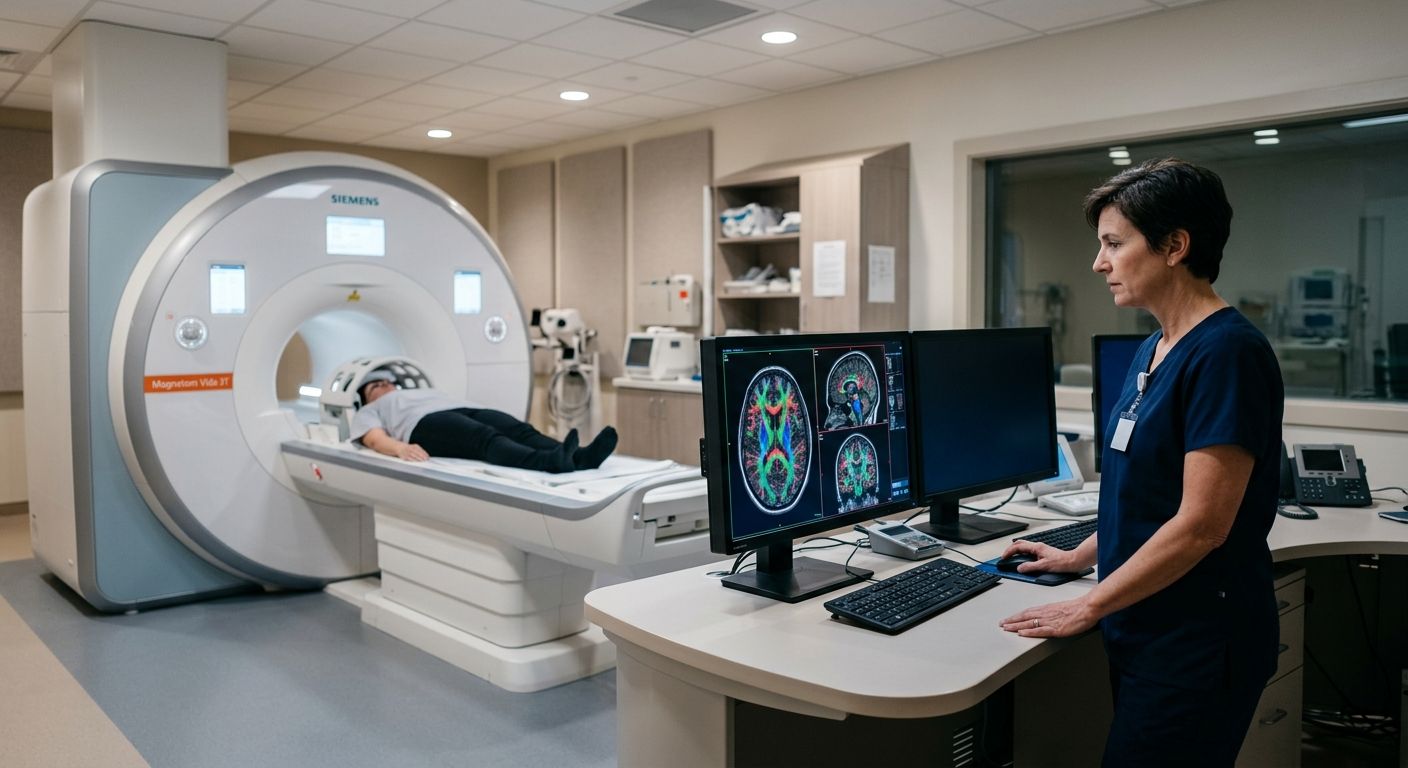
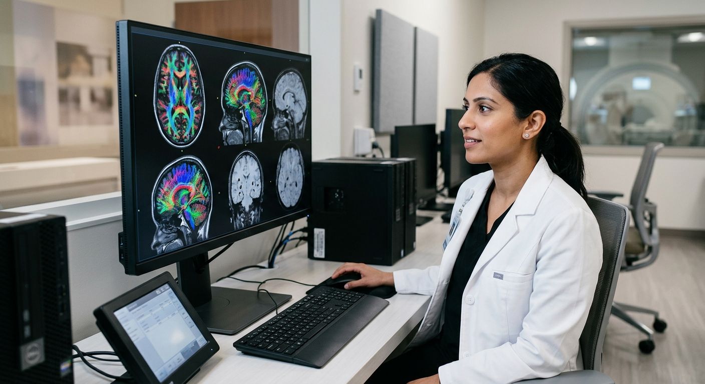
Board-certified radiologist reviewing advanced brain MRI scans on a large monitor in a modern imaging suite
Healthcare Legal Support, Medical Imaging Services, MRI & CT Diagnostics, Personal Injury Care, Radiology Protocols, Traumatic Brain Injury (TBI)

Traumatic Brain Injury Diagnostic Standards: DTI and SWI Insights

Table of Contents Understanding Traumatic Brain Injury Diagnostic Standards Clinical Frameworks and Guidelines for TBI Diagnosis Advanced MRI Techniques for TBI Detection Diffusion Tensor Imaging in Traumatic Axonal Injury Susceptibility Weighted Imaging and Microhemorrhage Detection Integrating Serum Biomarkers with Imaging Implementing TBI Diagnostic Imaging in Clinical and Legal Practice Coordinating with Physicians and Attorneys for Timely Imaging Streamlining Prior Authorizations and Liens for TBI Cases Ensuring Rapid Report Turnaround and Legal Documentation Biopsychosocial and Multimodal Approaches to TBI Assessment Frequently Asked Questions About TBI Diagnostic Standards Advancing TBI Care Through Accurate Diagnostic Imaging Understanding Traumatic Brain Injury Diagnostic Standards Understanding traumatic brain injury diagnostic standards is essential for accurate imaging and patient care. At Arizona Advanced Imaging LLC, we rely on proven protocols to detect both obvious and subtle brain injuries, guiding every step from initial assessment to comprehensive evaluation. Our board-certified neuroradiologists utilize standardized criteria to ensure clarity in every image and confidence in every decision. Computed tomography (CT) remains the first-line standard for acute TBI. Its speed in detecting hemorrhages and fractures makes it indispensable in emergency settings. For subacute and chronic evaluations, magnetic resonance imaging (MRI) offers superior resolution, revealing diffuse axonal injuries or small contusions that CT may miss. We adhere to these established standards because timely, accurate diagnosis directly shapes treatment planning and recovery outcomes. Emerging advanced techniques, such as diffusion tensor imaging and susceptibility weighted imaging, are refining traumatic brain injury diagnostic standards. DTI maps white matter tracts to detect microstructural damage, while SWI heightens sensitivity to minute hemorrhages. Our fellowship-trained specialists employ these modalities when standard imaging proves inconclusive, ensuring no injury remains hidden. Validated scoring systems and structured reporting further strengthen the medicolegal value of every study, supporting both clinical decisions and legal documentation. These standards evolve with injury severity and time since injury. For a broader overview, explore the patient guide to medical imaging. Please consult with a healthcare professional for diagnosis and treatment advice, as results may vary based on individual patient conditions and imaging quality. Clinical Frameworks and Guidelines for TBI Diagnosis Having examined the fundamentals of TBI diagnosis, we now compare the major clinical frameworks shaping current assessment. Current traumatic brain injury diagnostic standards draw from three complementary models, each informing the way we structure our TBI care imaging protocols. As Your Trusted Radiology Partner, we apply these evidence-based frameworks to every TBI evaluation. The 2026 AAN/CDC guidelines standardize diagnosis across care settings, relying on CT for acute triage and MRI for follow-up, with serum biomarkers S100B, GFAP, and UCH-L1 guiding imaging decisions. According to RadiologyInfo, a patient education resource from RSNA and ACR, CT remains first-line for acute head trauma assessment. The CBI-M framework classifies injury severity for targeted treatment using advanced MRI, including diffusion tensor imaging and susceptibility weighted imaging, to detect microstructural injuries like diffuse axonal injury that conventional scans frequently miss. Our Spin Tech MRI protocol employs these same advanced sequences, delivering quantitative DTI data and automated microbleed detection that directly support the CBI-M classification system and its treatment stratification approach. The four pillars of modern assessment integrate clinical examination, advanced imaging, biomarker analysis, and outcome tracking into a unified diagnostic approach. Beyond DTI and susceptibility weighted imaging, this model incorporates functional MRI for evaluating brain activity patterns following injury. The biomarker panel expands to include neurofilament light alongside GFAP and UCH-L1, enabling broader assessment of neuronal damage and recovery. Notably, the CBI-M framework does not explicitly define biomarker use, unlike both the 2026 guidelines and the four-pillar model. The following table provides a side-by-side comparison of these three diagnostic frameworks: Comparison of TBI Diagnostic Frameworks 2026 Clinical Guidelines CBI-M Framework Four Pillars of Modern Assessment Standardize diagnosis across care settings Classify injury severity for targeted treatment Integrate clinical, imaging, biomarker, and outcome data CT for acute triage, MRI for follow-up Advanced MRI (DTI/SWI) for microstructural injury DTI, SWI, functional MRI for comprehensive evaluation S100B, GFAP, UCH-L1 recommended Not explicitly defined GFAP, UCH-L1, neurofilament light integrated Advanced MRI Techniques for TBI Detection Diffusion Tensor Imaging in Traumatic Axonal Injury Diffusion tensor imaging measures the directional movement of water molecules along white matter tracts in the brain. In healthy tissue, water diffuses predictably along axonal bundles, but when axons are sheared or damaged, this organized diffusion becomes disrupted. The result is a measurable reduction in fractional anisotropy, a metric that reflects white matter integrity. As a fellowship-trained team, we at Arizona Advanced Imaging use quantitative DTI to detect these diffuse axonal injuries, which are often invisible on conventional MRI sequences. Our spin tech MRI TBI protocol incorporates DTI tractography to map white matter pathways and identify regions of reduced connectivity. Even in mild TBI cases where patients report persistent cognitive symptoms, susceptibility weighted imaging and diffusion tensor imaging can reveal objective evidence of injury that would otherwise go undetected. Susceptibility Weighted Imaging and Microhemorrhage Detection Susceptibility weighted imaging exploits magnetic susceptibility differences between tissues to detect blood products that accumulate after traumatic shearing forces tear small vessels. SWI is particularly effective at visualizing microhemorrhages and hemosiderin deposits that appear as tiny dark foci scattered throughout the brain parenchyma. Comparison of DTI and SWI for TBI Detection Comparison of DTI and SWI for TBI Detection Diffusion Tensor Imaging (DTI) Susceptibility Weighted Imaging (SWI) Measures water diffusion along white matter tracts Detects magnetic susceptibility differences from blood products Reduced fractional anisotropy indicating axonal injury Microhemorrhages, hemosiderin deposits High sensitivity for diffuse axonal injury Moderate sensitivity; better for hemorrhagic shear injuries Prognostic biomarker for cognitive outcome Identifies lesion burden correlating with severity DTI and SWI comparison for advanced TBI detection Integrating Serum Biomarkers with Imaging Emerging blood-based biomarkers offer an additional layer of diagnostic information that complements advanced MRI findings. After traumatic brain injury, proteins such as glial fibrillary acidic protein, ubiquitin C-terminal hydrolase L1, and neurofilament light are released from damaged neural cells into the bloodstream. Implementing TBI Diagnostic Imaging in Clinical and Legal Practice Coordinating with Physicians and Attorneys for Timely Imaging Successful TBI case

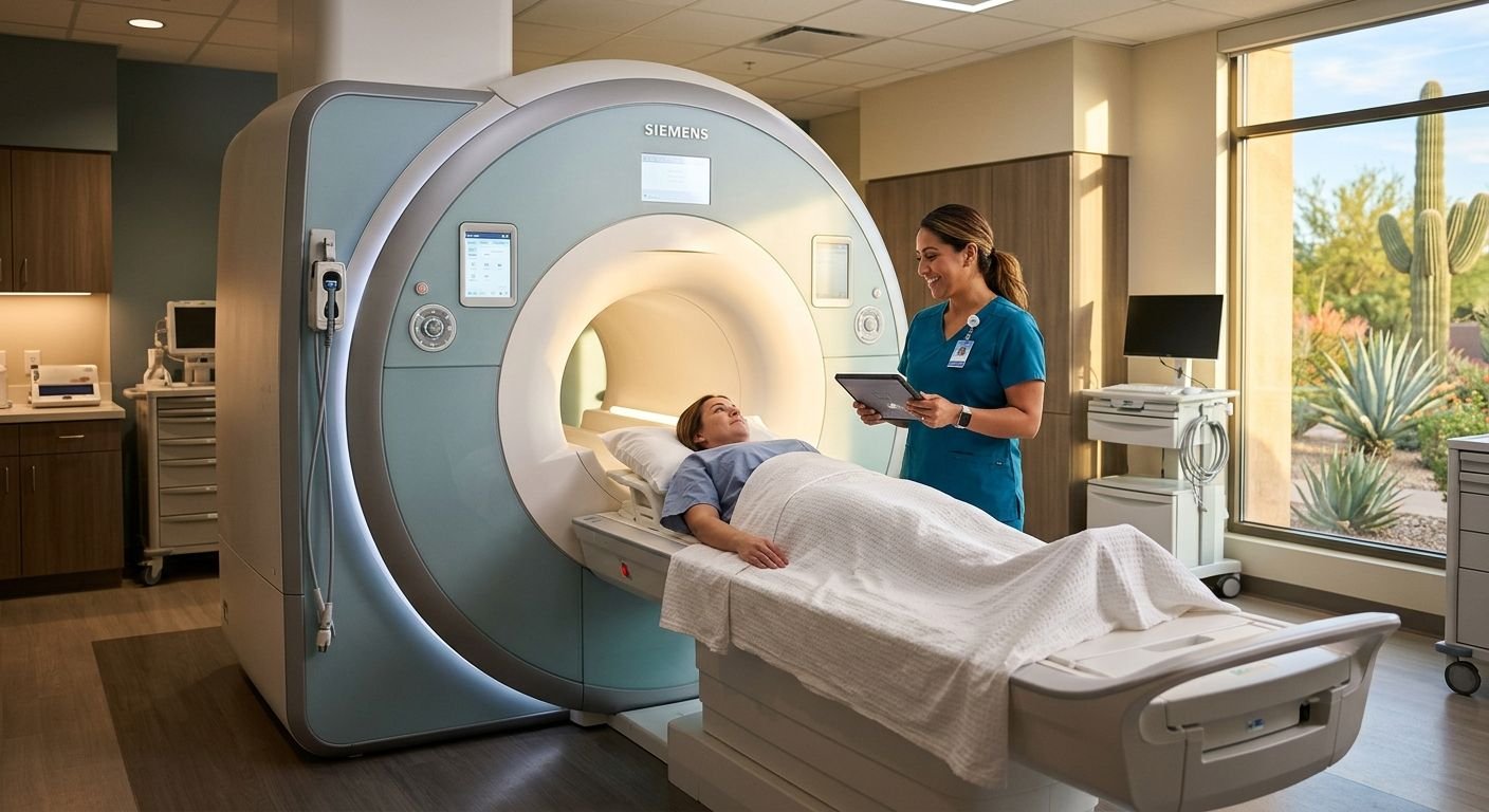
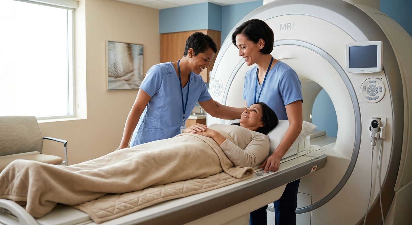
Compassionate imaging technologist assisting patient in modern MRI suite with open MRI machine
Diagnostic Imaging, Health & Wellness, Legal & Medical Coordination, Medical Facilities, MRI & CT Scans, Patient Education, Personal Injury Care, Radiology Services

Diagnostic Imaging for Personal Injury: MRI Guide in Phoenix

Table of Contents Diagnostic Imaging for Personal Injury and Orthopedic Care Understanding Diagnostic Imaging Modalities MRI: Magnetic Resonance Imaging for Soft Tissue Detail CT Scan: Advanced Cross-Sectional Imaging Ultrasound and X-Ray: Versatile Diagnostic Tools Benefits of Choosing a Specialized Imaging Center Fellowship-Trained Radiologists for Accurate Diagnoses Rapid Turnaround and Legal Coordination Convenient Locations and Concierge Services How Diagnostic Imaging Works at Our Centers Scheduling and Preparation The Imaging Procedure Report Generation and Digital Delivery Best Practices for Coordinating Imaging with Legal and Medical Teams Obtaining Court-Ready Radiology Reports Managing Billing with Letters of Protection Selecting the Right Imaging Center for Your Case Partnering with Arizona Advanced Imaging for Reliable Results Diagnostic Imaging for Personal Injury and Orthopedic Care Now, let’s explore the specific diagnostic imaging options available for personal injury and orthopedic care. When accidents occur, these advanced tools provide the clarity needed for accurate diagnosis and effective treatment planning, directly supporting both your recovery and any associated legal claims. MRI for soft tissue injuries: A personal injury MRI excels at revealing soft tissue damage invisible on X-rays. We use it to detect ligament tears, herniated discs, and muscle strains, which are common after accidents. Its precise imaging is vital for documenting these injuries thoroughly. CT scans for complex trauma: For detailed bone fracture evaluation and detecting traumatic brain injuries (TBI), CT scans are indispensable. They provide a rapid, comprehensive view of the skull, spine, and skeletal system after significant trauma. X-ray for initial assessment: Serving as a quick, first-line tool, X-rays efficiently identify obvious fractures and dislocations, giving us an immediate insight into your orthopedic condition. Ultrasound for soft tissue issues: Musculoskeletal radiology extends to ultrasound, a safe, radiation-free method ideal for assessing conditions like tendonitis and bursitis, offering dynamic evaluation where needed. Timely, same-day or next-day appointments are critical for documenting injury proximity to an accident. All studies are reviewed by fellowship-trained radiologists, with a report turnaround within 24–48 hours, supporting prompt clinical decisions and legal needs. Your health and clarity are our priority. With these imaging capabilities, we are ready to provide the clarity you need. Learn more or schedule an appointment for advanced imaging in Arizona contact. Please consult with a healthcare professional for diagnosis; radiation risks may apply. Results may vary. Understanding Diagnostic Imaging Modalities Understanding diagnostic imaging modalities helps patients and providers choose the right tool for each case. Our practice offers four core scanning methods, each designed to capture the precise detail needed for accurate diagnosis and effective personal injury care. MRI: Magnetic Resonance Imaging for Soft Tissue Detail Magnetic resonance imaging (MRI) uses strong magnetic fields and radio waves to create highly detailed images of soft tissues without any ionizing radiation. This makes it ideal for evaluating the brain, spinal cord, muscles, ligaments, and joints. In traumatic brain injury (TBI) cases, MRI can detect subtle abnormalities that other modalities might overlook. As a cornerstone of musculoskeletal radiology, MRI provides precise visualization of tendon tears, cartilage damage, and complex joint structures. For more detailed information, patients can refer to our radiology patient education resources. In personal injury MRI cases, the clarity of these scans supports both medical treatment and legal documentation. Our board-certified radiologists deliver reports within 24 to 48 hours, ensuring that attorneys and physicians have the evidence they need without delay. CT Scan: Advanced Cross-Sectional Imaging Computed tomography (CT) uses X-rays to generate cross-sectional images of the body, often in just minutes. This speed makes CT especially valuable for acute trauma and bone injuries. At Arizona Advanced Imaging, our same-day and next-day CT appointments help personal injury patients receive fast, accurate assessments after motor vehicle accidents or workplace incidents. The modality excels at detecting fractures, internal bleeding, and spinal damage, and our CT angiography studies provide detailed vascular imaging for complex cases. While CT involves a small dose of radiation, modern equipment with dose-reduction technology keeps exposure as low as possible. Because CT and MRI serve different purposes, our fellowship-trained radiologists select the optimal study for each injury—CT when speed and bone detail are paramount, and MRI when soft-tissue evaluation is needed. Ultrasound and X-Ray: Versatile Diagnostic Tools Ultrasound uses high-frequency sound waves to produce real-time images of soft tissues and blood flow, completely free of ionizing radiation. For musculoskeletal evaluations, our technologists can dynamically assess tendons, muscles, and joints—observing how they move during an exam. Vascular Doppler studies extend this capability to blood flow analysis, helping detect clots or vessel narrowing after trauma. X-ray, by contrast, remains the go-to tool for rapid bone and chest imaging; it is often the first test ordered when fractures or dislocations are suspected. Each of these diagnostic imaging techniques plays a distinct role in personal injury care: ultrasound offers dynamic, radiation-free soft-tissue assessment, while X-ray provides immediate fracture screening. Both are available with walk-in convenience and our standard 24-to-48-hour report turnaround, reinforcing our commitment as Your Trusted Radiology Partner. Each modality serves a unique purpose, and our team ensures you get the right scan for your specific needs. We combine advanced technology with board-certified expertise so that every image delivers clarity in every image, confidence in every decision. Benefits of Choosing a Specialized Imaging Center Specialized diagnostic imaging for personal injury cases requires expertise beyond standard scans. At Arizona Advanced Imaging, our advanced imaging personal injury services are designed to support legal teams with fast, accurate results. We offer a seamless experience that combines expert interpretation, rapid legal coordination, and patient-centered convenience, ensuring every case is supported by clear, defensible evidence. Fellowship-Trained Radiologists for Accurate Diagnoses The quality of an imaging report is only as strong as the radiologist interpreting it. Our team includes board-certified, fellowship-trained specialists in neuroradiology and musculoskeletal radiology. This sub-specialty training is critical for personal injury cases, as it enables the precise detection of subtle, trauma-related conditions, such as traumatic brain injuries and complex ligament tears, that general radiologists might overlook. According to Arizona Advanced Imaging, our radiologists’ deep focus on trauma-related pathologies means their findings are both detailed

Scroll to Top