CBI-M Traumatic Brain Injury Framework: A Complete Guide
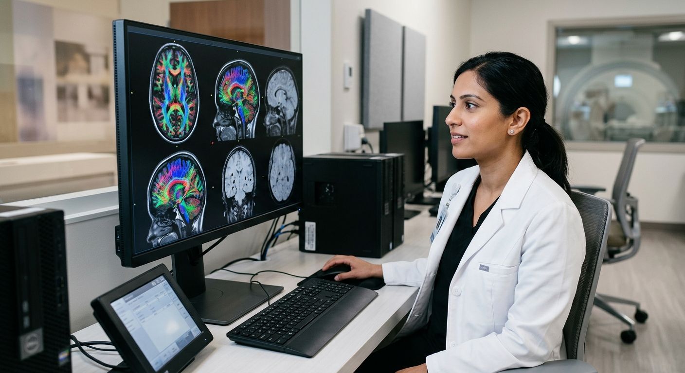
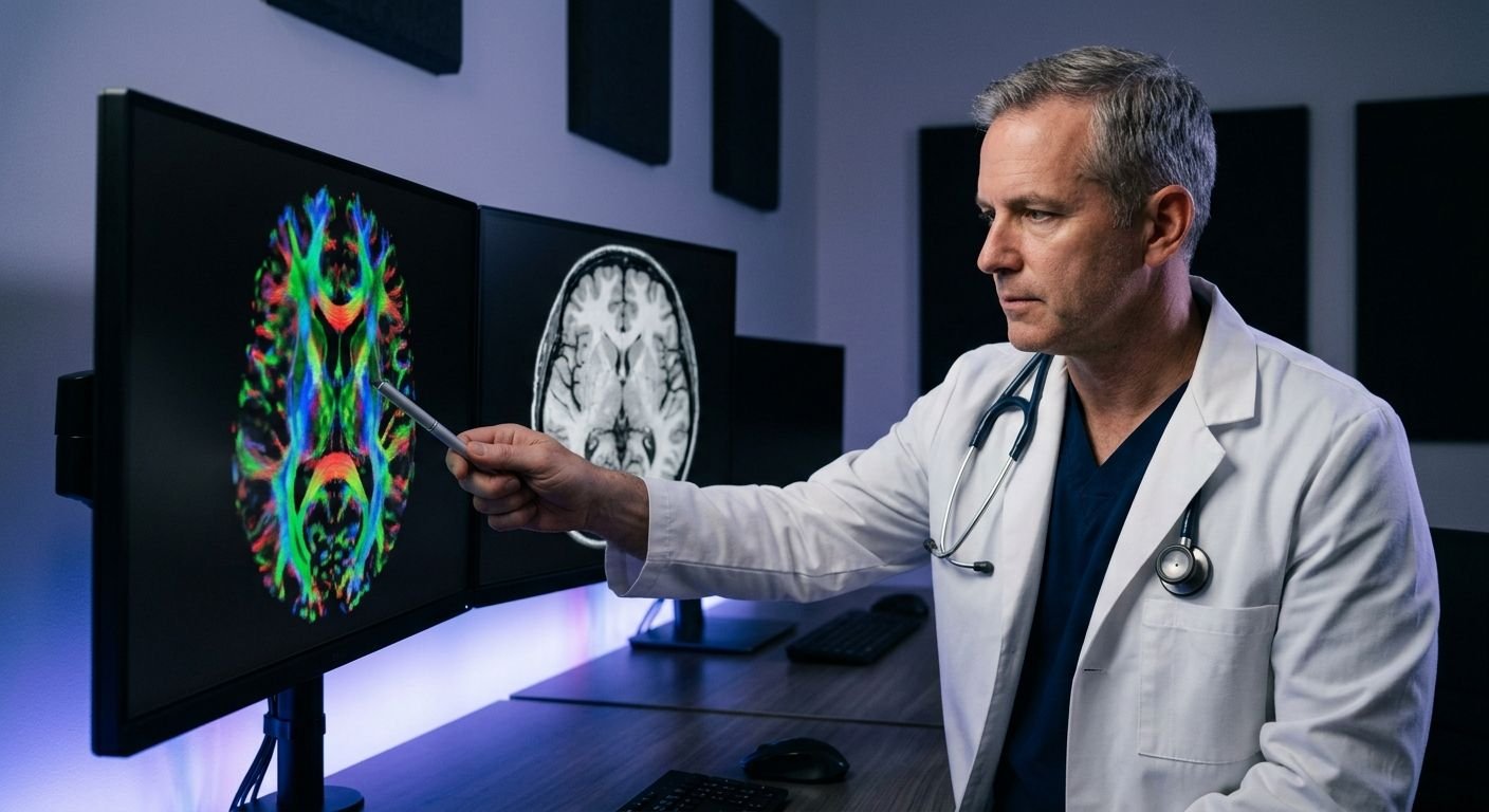
Table of Contents A Comprehensive Guide to the CBI-M Traumatic Brain Injury Framework Foundations of Traumatic Brain Injury Classification and the CBI-M Model Imaging and Biomarker Integration: The Core Pillar of the CBI-M Framework The Role of Advanced MRI and CT in CBI-M How Quantitative Imaging Enhances Mild TBI Diagnosis CBI-M vs. Glasgow Coma Scale: Why Multidimensional Wins Implementing the CBI-M Framework in Clinical and Legal Settings Streamlining Workflows for Personal Injury Cases Using DTI and SWI for Forensic‑Quality Reports Ensuring Rapid Turnaround and Court‑Ready Documentation Quantitative DTI and SWI: Deepening Diagnostic Precision Common Questions About CBI-M and TBI Imaging Elevating TBI Care with the CBI-M Framework A Comprehensive Guide to the CBI-M Traumatic Brain Injury Framework The CBI-M traumatic brain injury framework is a structured classification system that helps clinicians and imaging teams understand the type and severity of traumatic brain injuries. We rely on such classification to guide our imaging recommendations—from open MRI scans to specialized TBI protocols—so that physicians and legal teams receive reliable, objective data. We explain what the CBI-M framework is, how it organizes brain injury categories, and why that structure leads to more targeted imaging choices. We provide a basic overview of TBI classification, cover the key elements of the CBI-M model, and describe the application of advanced MR imaging protocols (such as the SpinTech™ MRI TBI Protocol) to identify microhemorrhages and alterations of white matter in the brain. This document is intended for the general audience, e.g. patients and/or their family members evaluating their imaging options, and/or attorneys and law firms evaluating potential personal injury claims. We acknowledge the importance of clinical and/or subjective assessment and complementary imaging in the evaluation of TBI. Our commitment to clarity in every image and confidence in every decision flows from this classification-first mindset. Therefore, in line with our classification-first strategy, we will cover the basic elements of TBI classification and the place of CBI-M before describing the imaging options. Foundations of Traumatic Brain Injury Classification and the CBI-M Model Traditionally, traumatic brain injuries (TBIs) are classified as mild, moderate, or severe, often using the Glasgow Coma Scale (GCS) to measure severity. A patient scores mild TBI when their GCS score is 13 to 15. A patient scores moderate TBI when their GCS score is 9 to 12. A patient scores severe TBI when their GCS score is 8 or less. Duration of loss of consciousness and length of post-traumatic amnesia are added to sharpen the severity picture. Two patients with the same GCS score may show different symptoms, injury patterns, and recovery rates. The majority of TBIs are classified as mild. Often, conventional Computed Tomography (CT) scans or Magnetic Resonance Imaging (MRIs) do not show abnormalities, and patients are left to manage persistent post-concussive symptoms. Trying to fill that gap, the CBI-M model uses a multi-pillar assessment instead of a single assessment. The four pillars of the model are: Clinical History, Symptom Burden, Functional Assessment, and Imaging & Biomarkers. CBI-M Pillars vs. Traditional TBI Assessment Assessment Component CBI-M Framework Traditional Approach Clinical Advantage Clinical History Detailed accident history, prior concussions, and comorbidities Loss of consciousness duration Adds context beyond a single acute variable Symptom Burden Type, severity, and persistence of symptoms via validated checklists Post-traumatic amnesia duration Reflects daily patient experience, not just one parameter Functional Assessment Cognitive, balance, and oculomotor testing post-injury Glasgow Coma Scale Detects subtle functional deficits GCS cannot measure Imaging & Biomarkers Advanced MRI (quantitative DTI, volumetric analysis, microbleed detection) Conventional CT or MRI Reveals microstructural injury invisible to routine scans Functional assessment may uncover balance or processing-speed deficits that significantly affect daily life while routine imaging appears unchanged. The Imaging & Biomarkers pillar serves as the model’s objective anchor. Our centers utilize the SpinTech MRI TBI protocol, an advanced imaging sequence, which incorporates quantitative diffusion tensor imaging, and is able to detect microstructural changes that may be missed in standard sequences. As your trusted radiology partner, we provide this protocol throughout Arizona and Texas. Reports, reviewed by a fellowship-trained neuroradiologist, are provided within 24-48 hours. CBI-M Pillars versus traditional concussion assessment compared side by side Radiology, therefore, becomes the objective, quantifiable anchor for clinical and symptom burden and functional performance. For patients and their referring clinicians, we encourage imaging finding review with a qualified physician to aid in placement of the imaging findings in the context of symptoms and to assist with care planning. Imaging and Biomarker Integration: The Core Pillar of the CBI-M Framework The CBI-M framework is built upon objective, tissue-level evidence. Advanced imaging provides that layer. Unlike assessment tools that depend on patient self-report or bedside observation alone, imaging-derived biomarkers record injury directly from brain tissue. The Role of Advanced MRI and CT in CBI-M Standard emergency CT and conventional MRI protocols were not designed to capture the microstructural and microvascular injuries that define subacute and chronic traumatic brain injury. The CBI-M framework addresses this gap by integrating advanced MRI sequences, diffusion tensor imaging, or DTI, which measures the integrity of white-matter tracts by tracking water molecule movement, and susceptibility weighted imaging, which amplifies the magnetic signal of blood products to reveal microhemorrhages — along with CT when indicated. Our TBI-specific protocol exemplifies how dedicated imaging protocols strengthen the CBI-M evidence layer. The FDA-cleared equipment allows for quantitative DTI and volumetric analysis of the brain, as well as for the automated detection of microbleeds. Conventional MRI may miss up to 50 percent of cerebral microbleeds present in mild TBI cases, while this protocol is backed by more than 100 published research studies supporting its diagnostic sensitivity. Fellowship-trained, board-certified neuroradiologists interpret the studies and create structured reports across our network of centers in Arizona and Texas. How Quantitative Imaging Enhances Mild TBI Diagnosis The CBI-M framework alters the diagnostic equation to incorporate quantitative DTI (diffusion tensor imaging) metrics (i.e. fractional anisotropy and mean diffusivity) to describe the directional movement of water molecules along axonal pathways, as well as susceptibility-weighted imaging signal voids, which represent sites of parenchymal hemorrhage.