AZ & TX Advanced Imaging

Medical-Legal Services

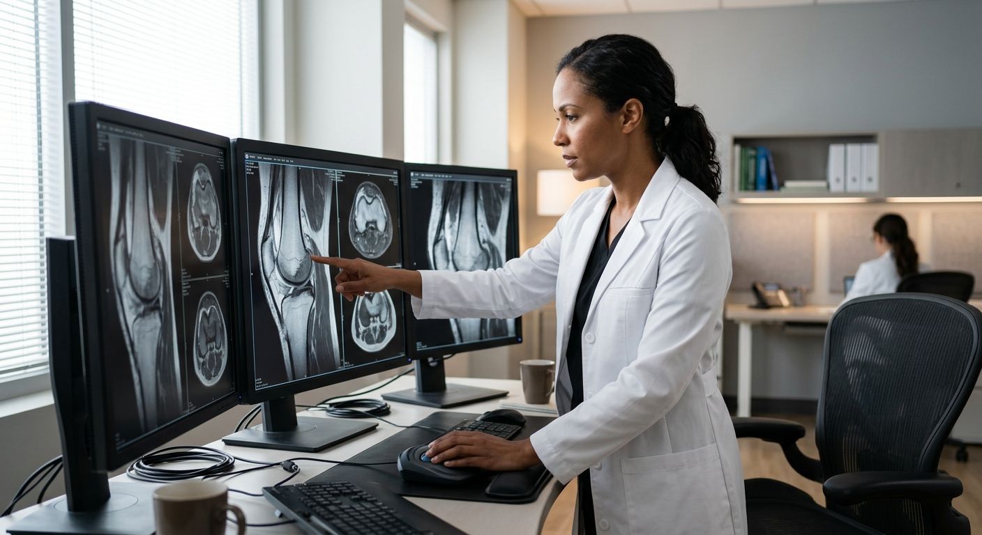
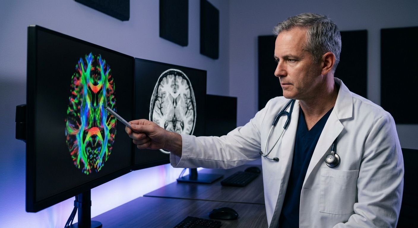
Board-certified radiologist analyzing high-resolution musculoskeletal MRI scans on multiple monitors in a modern medical reading room
Medical-Legal Services, MRI & CT Scans, Orthopedics

Top Musculoskeletal MRI Reviews: Phoenix Imaging Guide

Table of Contents Evaluating Musculoskeletal MRI Reviews with Confidence 1. Fellowship-Trained MSK Radiologists 2. Authentic Patient Reviews 3. Sports Injury Imaging Testimonials 4. Open MRI vs. Closed MRI 5. Rapid Report Turnaround 6. Detailed MSK Findings 7. Financial Ease with Liens and LOPs 8. Same-Day Appointment Access Select a Provider That Prioritizes Clarity and Speed Evaluating Musculoskeletal MRI Reviews with Confidence When evaluating musculoskeletal MRI reviews, confidence begins with understanding who interprets your imaging. At Arizona Advanced Imaging in Phoenix and Texas, our board-certified, fellowship-trained radiologists specialize in correlating musculoskeletal MRI findings with clinical history, ensuring each personal injury radiology report provides detailed, actionable insights. We deliver case-ready reports within 24 to 48 hours, allowing attorneys and physicians to make timely decisions without delay. Our reports prioritize clarity, combining annotated images with thorough descriptions so every stakeholder can proceed with certainty. We also accept liens and letters of protection, removing financial barriers for personal injury patients. For evaluations you can rely on, turn to our personal injury radiology services. 1. Fellowship-Trained MSK Radiologists Our team includes fellowship-trained MSK radiologists, specialists who have completed an additional one to two years of subspecialty training focused on musculoskeletal imaging after completing a full diagnostic radiology residency and achieving board certification. According to the American College of Radiology, this advanced training ensures deep expertise in interpreting MRI, CT, ultrasound, and X-ray examinations of bones, joints, and soft tissues. The quality of musculoskeletal MRI reviews depends heavily on the radiologist’s training, which is why our specialists are uniquely qualified to identify subtle abnormalities that general radiologists might overlook. For personal injury cases, the accurate interpretation of musculoskeletal MRI findings can make a decisive difference in both treatment planning and legal outcomes. Our board-certified, fellowship-trained specialists bring unparalleled precision to personal injury diagnostic imaging, where every personal injury radiology report we produce must withstand rigorous scrutiny. Our radiologists follow strict guidelines set by the American College of Radiology, though individual results may vary. This commitment to excellence ensures that referring physicians and attorneys in Phoenix and across Arizona receive the clarity and confidence they need to move cases forward. 2. Authentic Patient Reviews Beyond our advanced technology, our patients’ experiences speak for themselves. Our musculoskeletal MRI reviews consistently highlight the convenience and clarity we provide in Phoenix, from same-day scheduling to rapid report delivery. One patient shared, “After my accident, Arizona Advanced Imaging scheduled my MRI the same day and delivered a detailed report within 24 hours. The musculoskeletal MRI findings were so precise that my attorney could immediately move forward with my case—plus they handled the lien paperwork for me.” Another patient noted, “The walk-in ease and multilingual support made a stressful time much easier. My personal injury radiology report was thorough and court-ready, which gave me confidence in my legal and medical journey.” A third patient added, “I appreciated how the staff explained my musculoskeletal MRI findings in Spanish, ensuring I fully understood my diagnosis. Their attention to detail and the speed of service made a real difference.” These patient stories reflect our commitment to clarity and care—a dedication that extends through every step of your visit. Your health and clarity are our priority — discover why our patients trust us. 3. Sports Injury Imaging Testimonials “I tore my ACL during a weekend game and needed answers fast. Arizona Advanced Imaging got me in the next day, and the musculoskeletal MRI findings gave my orthopedic specialist exactly what he needed to plan my surgery. The images were incredibly sharp, and the personal injury radiology report was court-ready in under 48 hours. I felt supported every step of the way—they really are Your Trusted Radiology Partner.” — Mark, Phoenix athlete With over two decades of PI-focused radiology expertise, we combine 24–48 hour report turnaround with the clarity of board-certified, fellowship-trained radiologists. Ready to experience the same clarity and confidence? Contact us to schedule your appointment. For more details on what to expect during your scan, visit our patient radiology information resource. Results may vary based on individual patient conditions. Please consult with a healthcare professional for diagnosis and treatment advice. 4. Open MRI vs. Closed MRI Both open and closed MRI systems serve distinct diagnostic purposes. For patients exploring musculoskeletal mri reviews, understanding these differences is crucial: closed MRI units typically operate at 1.5T to 3.0T, delivering the high-resolution images essential for detailed musculoskeletal mri findings, while open MRI scanners, often ranging from 0.2T to 0.7T, prioritize patient comfort with a wider, less confining design that accommodates larger body types and reduces claustrophobia. For personal injury cases, closed MRI remains the standard for comprehensive orthopedic and neurological assessment because its superior image clarity supports a thorough personal injury radiology report. We offer both options at Arizona Advanced Imaging in Phoenix, and our team recommends the most appropriate scanner based on your specific condition and legal documentation needs. Your health and clarity are our priority. Results may vary based on individual patient conditions. Please consult with a healthcare professional for diagnosis and treatment advice. Closed MRI vs. Open MRI – key differences and trade-offs 5. Rapid Report Turnaround Beyond expert interpretation, we also prioritize timely delivery of our findings through our 24 – 48 Hour Report Turnaround. Rapid report turnaround directly influences musculoskeletal MRI reviews in Phoenix by supporting faster case resolution. Our standard turnaround for finalized radiology reports is 24 to 48 hours, a timeline that can significantly affect the direction of personal injury cases. When reports are completed quickly, attorneys gain earlier clarity on liability and damages, allowing them to negotiate settlements from a position of strength. Treating physicians can initiate or adjust care without unnecessary delays, improving patient outcomes. Each personal injury radiology report includes detailed musculoskeletal MRI findings and is structured to meet the rigorous demands of courtroom evidence. Despite the speed, diagnostic accuracy is never compromised: every study is interpreted by the same board-certified, fellowship-trained radiologists who uphold our standard of precision. Experience our rapid turnaround firsthand through our personal

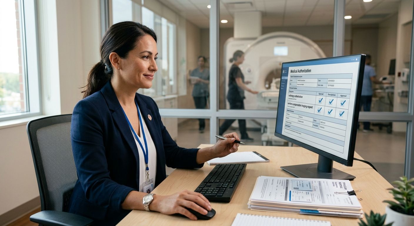
A medical authorization coordinator reviewing a workers' compensation imaging request on a computer screen in a modern office with diagnostic equipment visible through a window
Legal & Medical Coordination, Medical-Legal Services, Patient Resources

Workers Compensation Radiology Authorization: Process Explained

Table of Contents Workers’ Compensation Radiology Authorization in Arizona and Texas: A Step-by-Step Guide What You Need Before Requesting Imaging Authorization for Workers’ Comp in Arizona and Texas Understand the Workers’ Comp Radiology Authorization Process Gather Required Clinical Documentation for Workers’ Comp Imaging Verify Insurance Coverage and Pre-authorization Requirements Submit the Authorization Request with Medical Necessity Justification Follow Up and Track Your Authorization Status Handle Denials and Appeal Utilization Review Decisions Schedule Your Imaging Appointment After Approval Common Authorization Delays and How to Resolve Them Navigating Workers’ Comp Radiology Authorization with Confidence Workers’ Compensation Radiology Authorization in Arizona and Texas: A Step-by-Step Guide The workers compensation radiology authorization process ensures injured employees in Arizona and Texas receive necessary imaging while meeting carrier requirements. As Your Trusted Radiology Partner, we guide patients through each stage of the workers’ comp imaging approval pathway. Physician order and request submission. The treating doctor orders an MRI or CT scan and sends the request to the workers’ comp carrier or its utilization review organization. Radiology utilization review. A medical professional evaluates the order against evidence-based guidelines such as the ACR Appropriateness Criteria to confirm the radiology utilization review process supports the exam. Medical necessity determination. The reviewer confirms medical necessity for workers compensation MRI by verifying that the injury is work-related, conservative care has failed, and specific clinical findings justify advanced imaging. Authorization and scheduling. Once approved, an authorization number is issued. We coordinate your appointment and handle insurance and pre-authorization details so you can schedule with confidence. Workers’ compensation authorization rules differ by state and carrier; always check with your adjuster or provider. For a deeper understanding of imaging procedures, explore the patient guide to radiology. Contact AZ Advanced Imaging for personalized assistance with your workers’ comp imaging needs. What You Need Before Requesting Imaging Authorization for Workers’ Comp in Arizona and Texas Proper documentation is the cornerstone of a smooth workers compensation radiology authorization in Arizona and Texas. Before any imaging study can proceed, the referring physician and the patient must gather several key items. A written referral must be issued by the treating physician, specifying the imaging modality (MRI, CT, etc.), the anatomic area, ICD-10 diagnosis codes, and a concise clinical rationale. Next, proof of the work-related injury is required: the workers’ compensation claim number, date of injury, employer name, and the insurance carrier’s contact information. Additionally, documentation that establishes medical necessity for workers compensation MRI is essential—this typically includes progress notes, physical exam findings, and records of failed conservative treatment. The radiology utilization review process then kicks in: the workers’ comp carrier must approve the study, often after our team submits a utilization review request with all supporting records. State-specific rules in Arizona and Texas, such as Texas non-subscriber versus subscriber requirements, may also apply. At AZ Advanced Imaging, we verify and handle these forms, removing the administrative burden so patients and physicians can focus on care. Once these documents are ready, the authorization can advance smoothly, bringing clarity in every image and confidence in every decision. For comprehensive personal injury radiology services, including workers’ comp authorization support, contact us—your trusted radiology partner. Understand the Workers’ Comp Radiology Authorization Process To receive imaging under workers’ comp, you first need to understand the authorization process. Workers compensation radiology authorization is the insurance pre-approval required before an injured worker can receive an MRI, CT, or X-ray. This step ensures the scan is based on medical necessity for workers compensation MRI or other studies, meaning your provider must submit clinical documentation showing the imaging is essential for diagnosing or treating your workplace injury. The radiology utilization review process involves the insurance carrier evaluating that request, often using evidence-based guidelines to decide on approval. Denials can occur for reasons like incomplete paperwork, but our team at Arizona Advanced Imaging assists in compiling the required documentation to help streamline the submission. While authorization timelines typically range from 3 to 10 business days, understanding this process helps you set realistic expectations and avoid frustration during your recovery. Workers’ comp radiology authorization process in three steps We handle pre-authorization so you can focus on healing—that’s what patient-centric personal injury imaging truly means. With our support, we aim to make this necessary administrative step as smooth as possible, keeping your care on track. Gather Required Clinical Documentation for Workers’ Comp Imaging To ensure a smooth authorization process, patients must gather the following clinical documentation. While this section focuses on workers’ compensation imaging, patients needing personal injury X-ray services will find similar documentation requirements. We know that complete paperwork can expedite your workers compensation radiology authorization, and we are here to help at every step. Physician Referral/Order: This mandatory document must state the clinical indication and demonstrate medical necessity for workers compensation MRI. Claim Details: Provide your workers’ compensation claim number and the insurance carrier’s name for proper billing. Pre-Authorization Proof: Obtain the pre-certification document from your carrier to prevent processing delays. Identification: Bring a valid photo ID and your personal insurance card, if applicable. Supporting Medical Records: Include clinical notes and the initial injury report, as the radiology utilization review process often relies on this history. Incomplete documentation can delay your care, but our team will review your documents before we submit them for authorization. Once you have gathered these items, our experts can efficiently manage the next steps. Verify Insurance Coverage and Pre-authorization Requirements Confirming coverage and obtaining pre-authorizations can be time-consuming, so we manage these steps on your behalf. Our team works directly with commercial insurers, Medicare, and workers’ compensation carriers, navigating the radiology utilization review process to establish medical necessity. When a workers’ compensation MRI is ordered, we provide the clinical documentation required to support the request and work proactively to prevent unnecessary denials. By securing authorization before your appointment, we help you avoid surprise bills and treatment delays. We also accommodate patients through personal injury liens and letters of protection, and we verify benefits for most major plans, including Blue Cross Blue Shield, Cigna,

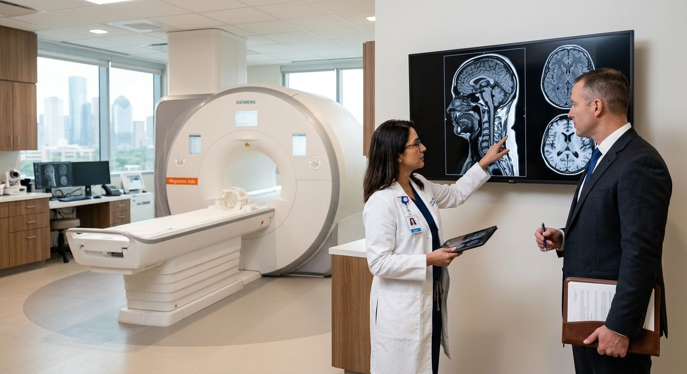
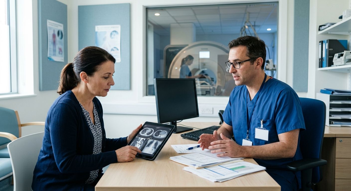
A female patient and a male radiologist in a modern radiology center discussing prior authorization, with an MRI machine in the background, no text visible.
Diagnostic Imaging, Medical-Legal Services, Patient Resources

Radiology Prior Authorization: Step-by-Step Guide for Providers

Table of Contents Radiology Prior Authorization: What Patients and Providers Need to Know 1. Understanding Prior Authorization in Radiology 2. How 2026 CMS Rules Impact Prior Authorization Timelines 3. Speeding Up Imaging Authorization for Injury Claims 4. Why Was Your MRI Prior Authorization Denied? 5. Documentation Required for Specialized Brain Imaging Approval 6. Can an Imaging Center Handle Pre-Authorizations for You? 7. Medical Necessity Criteria for MRI: A Closer Look 8. The Role of Peer-to-Peer Review in Radiology Simplifying Radiology Prior Authorization with the Right Partner Radiology Prior Authorization: What Patients and Providers Need to Know Radiology prior authorization is required by many insurers before covering advanced imaging like MRI and CT scans. According to CMS, prior authorization helps ensure appropriate use of comprehensive diagnostic imaging services. The authorization process typically follows four steps: Clinical documentation is submitted. Insurer reviews necessity. If denied, peer-to-peer review. Approval schedules imaging. Medical Necessity Criteria Insurers require that the ordered imaging meets specific medical necessity criteria for MRI or CT scans, as outlined by CMS guidelines. This includes a documented clinical reason and use of the appropriate modality. Peer-to-Peer Review If an initial authorization is denied, a peer-to-peer review radiology consultation lets the referring physician discuss the case with the insurer’s medical reviewer. This often resolves denials. We handle pre-authorization and accept personal injury liens and letters of protection—no upfront costs. Contact us for help. Results may vary; consult your healthcare professional for diagnosis and treatment advice. 1. Understanding Prior Authorization in Radiology Radiology prior authorization is a health plan requirement to approve imaging like MRI and CT before they are performed, ensuring medical necessity. Insurers use prior authorization to control costs, reduce unnecessary imaging, and ensure appropriate use of radiology resources. CMS sets medical necessity criteria that commercial insurers often follow. When a physician orders imaging, the referral center submits clinical information to the insurer. The request is reviewed against medical necessity criteria for MRI and other modalities. If denied, a peer-to-peer review radiology discussion between the ordering physician and an insurance medical director may be pursued. At Arizona Advanced Imaging, we handle prior authorization paperwork and insurer coordination on our patients’ behalf. As a provider of comprehensive diagnostic radiology services, we streamline these steps to reduce administrative burdens. We are committed to being Your Trusted Radiology Partner, helping you access needed imaging. Understanding this foundational step is key before exploring how the process works in practice. 2. How 2026 CMS Rules Impact Prior Authorization Timelines Building on these challenges, the Centers for Medicare & Medicaid Services (CMS) has updated its Medicare payment regulations for 2026, directly impacting radiology prior authorization timelines. According to CMS, the final rule mandates standard prior authorization decisions within 14 calendar days and expedited decisions within 72 hours. This requirement applies specifically to Medicare Advantage plans, creating a binding obligation for insurers and providers to adhere to these strict timeframes. To facilitate compliance, the rule requires electronic prior authorization and interoperability standards, which accelerate data exchange and minimize manual processing. Standardized medical necessity criteria for MRI and peer-to-peer review radiology protocols further support timely, clinically-driven decisions. At Arizona Advanced Imaging, we have aligned our pre-authorization support and 24–48 hour report turnaround to these mandates. Your health and clarity are our priority, and we ensure rapid imaging access without administrative delays. We coordinate directly with insurers to meet these timelines, so patients receive the scans they need without delay. 3. Speeding Up Imaging Authorization for Injury Claims For injury patients, radiology prior authorization can stall critical imaging. We handle all insurance pre-authorizations—verifying medical necessity criteria for MRI and conducting peer-to-peer review radiology when required—so you can focus on recovery. We also provide rapid trauma x-ray imaging to document injuries immediately. Across our Arizona and Texas centers, we accept personal injury liens and letters of protection, removing upfront costs entirely. Our team manages radiology prior authorization from start to finish, including same-day or next-day scheduling for MRI, CT, and X-ray. We handle every administrative hurdle so you can get scanned quickly. We take the burden off your shoulders so you never have to navigate complex insurance bureaucracy alone. Board-certified, fellowship-trained radiologists typically deliver court-ready reports within 24 to 48 hours, giving your attorney clear evidence to support your claim. We coordinate directly with your healthcare providers and legal counsel to keep claims moving. Your health and clarity are our priority. 4. Why Was Your MRI Prior Authorization Denied? But what happens when that authorization is denied? Understanding radiology prior authorization denials helps you act. Here’s why denials happen and what you can do next. The most frequent reason denials occur is lack of medical necessity documentation—insurers enforce strict medical necessity criteria for MRI, requiring clear clinical evidence such as progress notes, physical examination findings, and justification why MRI is needed now. According to AHRA, the medical imaging management association, incomplete or outdated referral details—like missing CPT codes or an old office note—can also trigger rejection. Additionally, many personal-injury plans require prior conservative treatment (e.g., six weeks of physical therapy) before they’ll approve an MRI. If you receive a denial, a peer-to-peer review radiology consultation—where our radiologist discusses your case with the insurance medical director—can often resolve the issue. At Arizona Advanced Imaging, we assist with pre-authorization paperwork and facilitate these discussions to help you get the imaging you need. 5. Documentation Required for Specialized Brain Imaging Approval Securing radiology prior authorization for specialized brain imaging begins with a complete set of supporting documents. When you coordinate with your referring provider early, you avoid common bottlenecks and keep your diagnostic timeline on track. A physician referral or order is the foundational document that initiates every prior authorization request. Detailed clinical notes and patient history establish the medical necessity criteria for MRI, demonstrating that the exam is appropriate for your symptoms. Prior imaging reports or scans are often requested to provide comparison and avoid duplicate studies. Thorough, well-organized documentation often satisfies medical necessity criteria for MRI upfront and reduces the likelihood

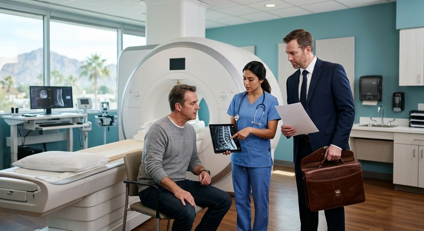
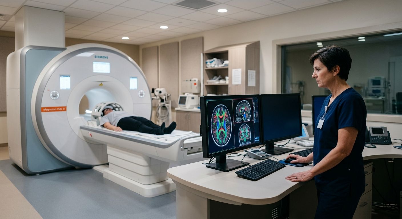
Modern MRI suite with advanced scanner and radiologist examining color-coded brain scan on monitor
Advanced MRI Technology, Diagnostic Imaging, Healthcare Technology, Medical-Legal Services, Patient Resources, Personal Injury Care, Traumatic Brain Injury (TBI)

Spintech MRI TBI Protocol: Complete Guide for Clinicians

Table of Contents The SpinTech MRI TBI Protocol: A New Standard in Brain Injury Detection How the SpinTech Protocol Detects Micro-Hemorrhages and Axonal Injury Exploring Quantitative Susceptibility Mapping and Diffuse Axonal Injury Detection The Role of Quantitative Susceptibility Mapping (QSM) Detecting Diffuse Axonal Injury with Advanced Sequences Accessing the SpinTech Protocol for TBI Imaging Across Arizona and Texas Finding Imaging Centers Equipped with the SpinTech Protocol What to Expect: Referral, Authorization, and Report Turnaround Coordinating with Legal Teams for Court-Ready Imaging Frequently Asked Questions About the SpinTech MRI TBI Protocol Advancing TBI Diagnosis with the SpinTech MRI Protocol The SpinTech MRI TBI Protocol: A New Standard in Brain Injury Detection Conventional MRI plays a critical role in brain imaging, yet it can miss up to half of the subtle microbleeds associated with mild traumatic brain injury. We address this diagnostic gap with the SpinTech MRI TBI protocol, a proprietary advanced imaging method available at AZ Advanced Imaging. By moving beyond standard structural scans, this approach provides objective evidence of injury that standard imaging often overlooks, helping patients and their physicians make informed decisions. This advanced TBI imaging protocol combines several specialized sequences to reveal what conventional scans cannot. Quantitative susceptibility mapping, or QSM, detects tiny deposits of iron from blood products, pinpointing microbleeds with exceptional sensitivity. Diffuse axonal injury detection is achieved through diffusion tensor imaging, or DTI, which assesses the integrity of white matter tracts and uncovers microstructural damage that disrupts normal brain communication. Susceptibility-weighted imaging, or SWI, further enhances the visualization of these otherwise invisible injuries. Every scan is interpreted by our board-certified, fellowship-trained radiologists, and we deliver a detailed report within 24 to 48 hours, ensuring you receive timely, actionable results. Clarity in every image, confidence in every decision. Our advanced TBI imaging protocol is particularly suited for personal injury patients who need objective and defensible documentation of brain injury. Results may vary based on individual patient conditions. We serve patients throughout Arizona and Texas from our primary location in Phoenix, providing access to technology that can make a decisive difference in both clinical care and legal proceedings. How the SpinTech Protocol Detects Micro-Hemorrhages and Axonal Injury Our spintech mri tbi protocol addresses the critical limitations of standard imaging by applying advanced sequences specifically designed to identify the subtle markers of mild traumatic brain injury. Where conventional scans may fail to detect up to 50% of cerebral microbleeds, this quantitative approach provides objective, measurable, and defensible results essential for both clinical treatment and legal documentation. Enhanced Detection of Micro-Hemorrhages The first pillar of the protocol’s diagnostic power lies in Susceptibility Weighted Imaging (SWI) and its advanced post-processing technique, quantitative susceptibility mapping. SWI is an MRI sequence exquisitely sensitive to hemosiderin, a blood breakdown product that accumulates in the brain after a micro-hemorrhage. On SWI images, these tiny bleeds appear as small, dark foci—findings that are often invisible on conventional scans. Our fellowship-trained, board-certified neuroradiologists then utilize QSM to go a step further, calculating the actual magnetic susceptibility of these lesions. This quantification allows for a crucial differentiation between acute and chronic micro-hemorrhages, mapping the full extent and age of the injury with a precision standard imaging cannot achieve. Identifying Diffuse Axonal Injury A second cornerstone of the SpinTech approach is diffuse axonal injury detection through Diffusion Tensor Imaging (DTI). While conventional MRI may show moderate sensitivity to this type of trauma, DTI provides a microscopic view of the brain’s white matter architecture. It works by measuring the directional diffusion of water molecules, which normally flow freely along intact axonal fiber tracts. When a shearing injury occurs—a hallmark of TBI from car accidents or falls—this orderly flow is disrupted. The protocol quantifies this disruption as a reduction in fractional anisotropy, a metric that serves as a direct, evidence-based indicator of axonal injury. This capability makes our protocol highly sensitive for detecting diffuse axonal injury that may otherwise go undiagnosed. The table below highlights key differences between a conventional brain MRI and our FDA-approved SpinTech protocol. Feature Conventional MRI SpinTech MRI TBI Protocol Ability to detect micro-hemorrhages Limited High sensitivity with SWI and QSM Scan time for brain 20-30 minutes 10-15 minutes (up to 50% faster) Sensitivity to diffuse axonal injury Moderate High with DTI and SWI Requirement for contrast agent Often needed Typically non-contrast By eliminating the need for a contrast agent in most cases and completing the scan in as little as 10 to 15 minutes, this non-contrast protocol significantly reduces patient risk and accelerates the diagnostic process. This combination of speed, safety, and sensitivity provides a clear advantage in building a comprehensive clinical picture. SpinTech MRI TBI Protocol vs. Conventional MRI comparison This integrated multi-sequence strategy is the foundation of our TBI diagnostic imaging legal support. By providing court-ready reports that objectively document the presence and nature of an injury, we give personal injury attorneys and physicians the clarity they need to make confident decisions for their cases and patients. Exploring Quantitative Susceptibility Mapping and Diffuse Axonal Injury Detection The Role of Quantitative Susceptibility Mapping (QSM) The SpinTech MRI TBI protocol represents an advanced imaging strategy that we employ to enhance the detection of subtle traumatic brain injuries. Building on the fundamental sensitivity of susceptibility weighted imaging, we now examine how quantitative susceptibility mapping (QSM) refines the identification of diffuse axonal injury. QSM is a post-processing technique that quantifies magnetic susceptibility within brain tissues, generating high-resolution maps that differentiate diamagnetic calcifications from paramagnetic blood products such as micro-hemorrhages. Detecting Diffuse Axonal Injury with Advanced Sequences Diffuse axonal injury detection relies on a multi-sequence approach that evaluates white matter integrity and micro-hemorrhage burden concurrently. Diffusion tensor imaging (DTI) is the primary tool for assessing the structural connectivity of white matter tracts, generating scalar metrics such as fractional anisotropy that decline when axons are sheared during acceleration-deceleration forces. Comparison of Diagnostic Sensitivity Technique Sensitivity to Micro-hemorrhages Specificity for DAI Clinical Utility Susceptibility Weighted Imaging (SWI) Very high Moderate Widely used, qualitative Quantitative Susceptibility Mapping (QSM) High High

Scroll to Top