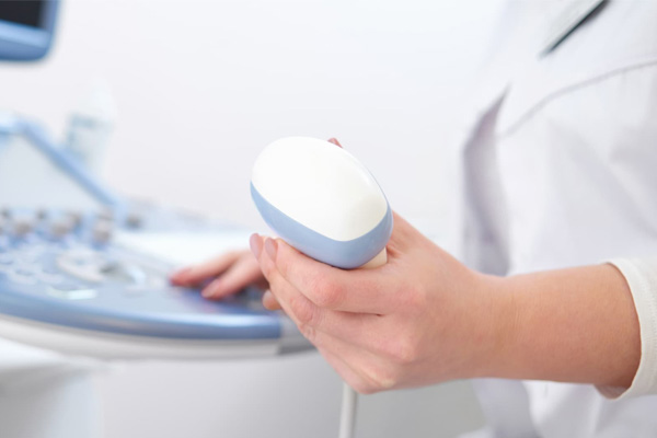
Ultrasound imaging (sonography) uses high-frequency sound waves to view inside the body. The ultrasound image is produced by the reflection of the waves off of the body structures. It is a combination of the time it takes for the sound signal to travel through the body as well as the strength of the signal as it is transmitted that creates the ultrasound image.
Because ultrasound images are captured in real-time, they can also show movement of the body’s internal organs as well as blood flowing through the blood vessels. This allows physicians to better evaluate function of body systems.
What to expect during an Ultrasound
You may be asked to wear a gown for your exam, depending on what part of your body will be examined.
In an ultrasound exam, a probe is placed directly on the skin or inside a body opening. A thin layer of gel is applied to the skin so that the ultrasound waves are transmitted from the transducer probe through the gel, into the body.
Typically, you will be able to see the screen that the Technologist is looking at, and you may have questions about what you are seeing, but as with any scan or test, a Radiologist must first review the images before information may be shared. You will receive your results from the doctor who ordered your test.


Benefits
- Unlike X-ray imaging, there is no ionizing radiation exposure associated with ultrasound imaging.
- This modality allows for evaluation of real time function of organs and systems in the body.
Risks
- Per the FDA, although ultrasound imaging is generally considered safe when used prudently by appropriately trained health care providers, ultrasound energy has the potential to produce biological effects on the body.
- Ultrasound waves can heat the tissues slightly. In some cases, it can also produce small pockets of gas in body fluids or tissues (cavitation). The long-term consequences of these effects are still unknown.

