
AZ & TX Advanced Imaging

Healthcare Technology

Radiologist in scrubs reviewing a high-resolution medical scan on a large encrypted tablet in a modern radiology workspace, with transparent data stream and lock overlays symbolizing HIPAA security.
Healthcare Legal Support, Healthcare Technology, Medical-Legal Services

HIPAA Secure Medical Imaging Delivery: Complete Compliance Guide

Table of Contents HIPAA Secure Medical Imaging Delivery for Personal Injury Cases HIPAA-Compliant Image Encryption Secure Patient Portal Access Digital Rights Management for Imaging Audit Trail and Access Logs Role-Based Access Control (RBAC) Encrypted Email Delivery for Reports Secure API Integration for Legal Portals Biometric Authentication for Image Viewing Redaction Tools for Protected Health Information Forensic Imaging and Chain-of-Custody Tracking Comparison of Secure Medical Imaging Delivery Tools Ensuring Secure Medical Imaging Delivery HIPAA Secure Medical Imaging Delivery for Personal Injury Cases For personal injury cases, hipaa secure medical imaging delivery is critical to protecting sensitive health information during discovery and litigation. At Arizona diagnostic imaging center, we comply with HIPAA regulations by using encrypted digital portals and secure upload methods that keep your imaging records confidential. The National Institutes of Health establishes the privacy and security standards we follow, ensuring that every image and report meets federal requirements for protected health information. We provide court-ready reports and secure digital copies directly to you and your attorney through our provider portal. Our digital infrastructure allows referring physicians and legal teams to access diagnostic images with controlled, role-based permissions, maintaining a clear chain of custody. When you upload your documents or request imaging records, the transmission is encrypted end-to-end, and we authenticate all access requests before releasing any protected data. With a 24–48 hour report turnaround, we deliver timely results without compromising data security. We accept liens and letters of protection, enabling you to receive necessary diagnostic scans with no upfront costs while your case proceeds. Eligibility for lien-based imaging is determined upon consultation. Clarity in every image, confidence in every decision. HIPAA-Compliant Image Encryption Building on the foundations of secure image delivery, we focus specifically on encryption as a critical technical safeguard for HIPAA secure medical imaging delivery. Under the HIPAA Security Rule, encryption protects ePHI from unauthorized access by rendering data unreadable without the correct decryption keys. While the rule does not mandate a specific algorithm, it designates encryption as an addressable implementation specification that most covered entities, including our Arizona and Texas imaging centers, adopt as a reasonable and appropriate measure for the secure delivery of medical images under HIPAA. For data at rest on our secure servers, we utilize AES-256 encryption, the gold standard adopted by federal agencies, including the national institutes of health, for protecting sensitive health information. When images are transmitted to referring physicians or accessed through patient portals, TLS 1.2 and 1.3 protocols establish encrypted channels that safeguard data in transit. According to the American Healthcare Radiology Administrators (AHRA), radiology practices should treat encryption as a foundational component of a mature medical imaging security program, integrating it alongside robust key management practices. We ensure encryption is applied across our multi-state network, enabling HIPAA-compliant medical image delivery whether images are shared within Phoenix or across state lines to Texas. Effective encryption key management remains a separate but essential companion to any encryption implementation, ensuring that only authorized personnel can access decryption credentials. This approach helps protect patient data and reduces risk without implying absolute immunity from all threats. Our commitment to these standards ensures that every image transmission supports informed clinical and legal decisions — embodying our belief that Clarity in every image, confidence in every decision begins with uncompromising data security. In the next section, we will explore the complementary HIPAA-compliant data security measures, such as access controls and audit trails, that further strengthen our overall protection framework. Secure Patient Portal Access Building on our commitment to rapid report turnaround, we deliver those finalized reports and your medical images through a fully HIPAA-compliant medical imaging delivery system. Our secure patient portal provides you with immediate, protected online access to your personal health information the moment your results are ready. This HIPAA secure medical imaging delivery method ensures that your sensitive data is shielded by advanced encryption and continuous security monitoring, giving you peace of mind whenever you log in. After your scan, our board-certified radiologists complete their analysis within our standard 24 to 48 hour report turnaround window, and the finalized report becomes available to you in the portal without delay. You can securely view your imaging studies and detailed radiology reports from any internet-connected device, empowering you to stay informed about your health on your own schedule. Our HIPAA-compliant medical imaging delivery framework follows stringent data privacy standards established by the American Society of Radiologic Technologists, reinforcing safe medical image access at every step. Beyond simply viewing your records, the patient portal enables seamless collaboration with your broader care team. You can electronically share your digital images and reports directly with referring physicians, specialists, or legal professionals, eliminating the delays and risks associated with traditional mail or fax. This protected online portal also serves as your permanent, centralized repository for all imaging performed at our centers, so your history is always organized and accessible when you need it. Our portal adheres to standards set by leading medical imaging management associations, reflecting our dedication to professional security and ethical data stewardship. We also follow guidance from the medical imaging management association to inform our protocols. Clarity in every image, confidence in every decision — that principle guides everything we build, including our digital patient experience. We designed this system to give you control over your diagnostic information while we maintain the strictest privacy protections. The next step after accessing your results is often a follow-up conversation or appointment, and our online scheduling tool makes that process just as convenient. Digital Rights Management for Imaging Building on our comprehensive imaging services, hipaa secure medical imaging delivery forms the foundation of how we protect and manage your diagnostic data. We define digital rights management in medical imaging as the integrated security protocols and access controls that safeguard patient images, ensuring only authorized parties can view or share sensitive health information. Unlike consumer-focused DRM tools, our approach centers entirely on clinical compliance and patient privacy. We maintain rigorous HIPAA compliance across every digital workflow, supported by encrypted
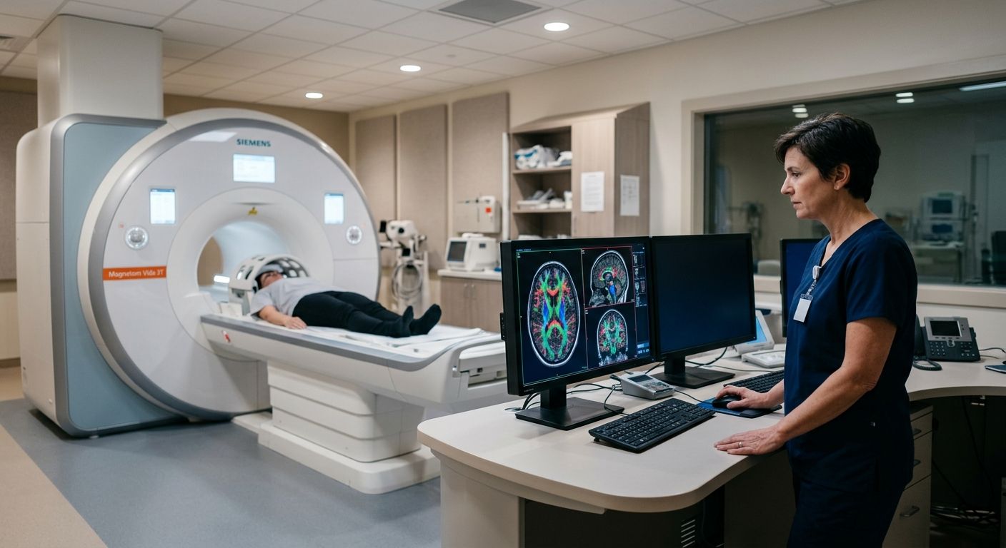
Modern MRI suite with advanced scanner and radiologist examining color-coded brain scan on monitor
Advanced MRI Technology, Diagnostic Imaging, Healthcare Technology, Medical-Legal Services, Patient Resources, Personal Injury Care, Traumatic Brain Injury (TBI)

Spintech MRI TBI Protocol: Complete Guide for Clinicians

Table of Contents The SpinTech MRI TBI Protocol: A New Standard in Brain Injury Detection How the SpinTech Protocol Detects Micro-Hemorrhages and Axonal Injury Exploring Quantitative Susceptibility Mapping and Diffuse Axonal Injury Detection The Role of Quantitative Susceptibility Mapping (QSM) Detecting Diffuse Axonal Injury with Advanced Sequences Accessing the SpinTech Protocol for TBI Imaging Across Arizona and Texas Finding Imaging Centers Equipped with the SpinTech Protocol What to Expect: Referral, Authorization, and Report Turnaround Coordinating with Legal Teams for Court-Ready Imaging Frequently Asked Questions About the SpinTech MRI TBI Protocol Advancing TBI Diagnosis with the SpinTech MRI Protocol The SpinTech MRI TBI Protocol: A New Standard in Brain Injury Detection Conventional MRI plays a critical role in brain imaging, yet it can miss up to half of the subtle microbleeds associated with mild traumatic brain injury. We address this diagnostic gap with the SpinTech MRI TBI protocol, a proprietary advanced imaging method available at AZ Advanced Imaging. By moving beyond standard structural scans, this approach provides objective evidence of injury that standard imaging often overlooks, helping patients and their physicians make informed decisions. This advanced TBI imaging protocol combines several specialized sequences to reveal what conventional scans cannot. Quantitative susceptibility mapping, or QSM, detects tiny deposits of iron from blood products, pinpointing microbleeds with exceptional sensitivity. Diffuse axonal injury detection is achieved through diffusion tensor imaging, or DTI, which assesses the integrity of white matter tracts and uncovers microstructural damage that disrupts normal brain communication. Susceptibility-weighted imaging, or SWI, further enhances the visualization of these otherwise invisible injuries. Every scan is interpreted by our board-certified, fellowship-trained radiologists, and we deliver a detailed report within 24 to 48 hours, ensuring you receive timely, actionable results. Clarity in every image, confidence in every decision. Our advanced TBI imaging protocol is particularly suited for personal injury patients who need objective and defensible documentation of brain injury. Results may vary based on individual patient conditions. We serve patients throughout Arizona and Texas from our primary location in Phoenix, providing access to technology that can make a decisive difference in both clinical care and legal proceedings. How the SpinTech Protocol Detects Micro-Hemorrhages and Axonal Injury Our spintech mri tbi protocol addresses the critical limitations of standard imaging by applying advanced sequences specifically designed to identify the subtle markers of mild traumatic brain injury. Where conventional scans may fail to detect up to 50% of cerebral microbleeds, this quantitative approach provides objective, measurable, and defensible results essential for both clinical treatment and legal documentation. Enhanced Detection of Micro-Hemorrhages The first pillar of the protocol’s diagnostic power lies in Susceptibility Weighted Imaging (SWI) and its advanced post-processing technique, quantitative susceptibility mapping. SWI is an MRI sequence exquisitely sensitive to hemosiderin, a blood breakdown product that accumulates in the brain after a micro-hemorrhage. On SWI images, these tiny bleeds appear as small, dark foci—findings that are often invisible on conventional scans. Our fellowship-trained, board-certified neuroradiologists then utilize QSM to go a step further, calculating the actual magnetic susceptibility of these lesions. This quantification allows for a crucial differentiation between acute and chronic micro-hemorrhages, mapping the full extent and age of the injury with a precision standard imaging cannot achieve. Identifying Diffuse Axonal Injury A second cornerstone of the SpinTech approach is diffuse axonal injury detection through Diffusion Tensor Imaging (DTI). While conventional MRI may show moderate sensitivity to this type of trauma, DTI provides a microscopic view of the brain’s white matter architecture. It works by measuring the directional diffusion of water molecules, which normally flow freely along intact axonal fiber tracts. When a shearing injury occurs—a hallmark of TBI from car accidents or falls—this orderly flow is disrupted. The protocol quantifies this disruption as a reduction in fractional anisotropy, a metric that serves as a direct, evidence-based indicator of axonal injury. This capability makes our protocol highly sensitive for detecting diffuse axonal injury that may otherwise go undiagnosed. The table below highlights key differences between a conventional brain MRI and our FDA-approved SpinTech protocol. Feature Conventional MRI SpinTech MRI TBI Protocol Ability to detect micro-hemorrhages Limited High sensitivity with SWI and QSM Scan time for brain 20-30 minutes 10-15 minutes (up to 50% faster) Sensitivity to diffuse axonal injury Moderate High with DTI and SWI Requirement for contrast agent Often needed Typically non-contrast By eliminating the need for a contrast agent in most cases and completing the scan in as little as 10 to 15 minutes, this non-contrast protocol significantly reduces patient risk and accelerates the diagnostic process. This combination of speed, safety, and sensitivity provides a clear advantage in building a comprehensive clinical picture. SpinTech MRI TBI Protocol vs. Conventional MRI comparison This integrated multi-sequence strategy is the foundation of our TBI diagnostic imaging legal support. By providing court-ready reports that objectively document the presence and nature of an injury, we give personal injury attorneys and physicians the clarity they need to make confident decisions for their cases and patients. Exploring Quantitative Susceptibility Mapping and Diffuse Axonal Injury Detection The Role of Quantitative Susceptibility Mapping (QSM) The SpinTech MRI TBI protocol represents an advanced imaging strategy that we employ to enhance the detection of subtle traumatic brain injuries. Building on the fundamental sensitivity of susceptibility weighted imaging, we now examine how quantitative susceptibility mapping (QSM) refines the identification of diffuse axonal injury. QSM is a post-processing technique that quantifies magnetic susceptibility within brain tissues, generating high-resolution maps that differentiate diamagnetic calcifications from paramagnetic blood products such as micro-hemorrhages. Detecting Diffuse Axonal Injury with Advanced Sequences Diffuse axonal injury detection relies on a multi-sequence approach that evaluates white matter integrity and micro-hemorrhage burden concurrently. Diffusion tensor imaging (DTI) is the primary tool for assessing the structural connectivity of white matter tracts, generating scalar metrics such as fractional anisotropy that decline when axons are sheared during acceleration-deceleration forces. Comparison of Diagnostic Sensitivity Technique Sensitivity to Micro-hemorrhages Specificity for DAI Clinical Utility Susceptibility Weighted Imaging (SWI) Very high Moderate Widely used, qualitative Quantitative Susceptibility Mapping (QSM) High High

Scroll to Top