AZ & TX Advanced Imaging

Medical Imaging Services

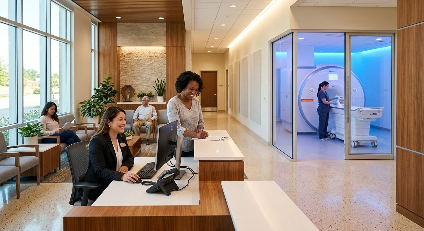
Interior of a modern freestanding MRI center in Texas with a smiling patient coordinator at the front desk and a hallway leading to a softly lit open MRI suite.
Diagnostic Imaging, Medical Facilities, Medical Imaging Services

Best Freestanding MRI Centers in Texas: Local Guide 2026

Table of Contents Freestanding MRI Centers in Texas for Reliable Diagnostic Imaging Comparing Freestanding MRI Centers Across Texas Markets Same-Day Appointments and Insurance Acceptance at Texas Freestanding Centers What Sets Freestanding MRI Centers Apart from Hospital Outpatient Departments How to Schedule Your MRI at a Freestanding Center in Texas Clarity and Confidence with Freestanding MRI Centers in Texas Freestanding MRI Centers in Texas for Reliable Diagnostic Imaging We bring the same patient-first approach that has defined our diagnostic imaging in Arizona to Texas, where our freestanding MRI centers provide efficient, compassionate care. Each facility offers MRI services. If you experience claustrophobia or need a particular scanner type, contact your preferred Texas center to confirm its scanner options before booking. We schedule same-day and next-day appointments to support urgent diagnostic needs. Every scan is reviewed by board-certified, fellowship-trained radiologists, and we consistently deliver detailed reports within 24 to 48 hours. We accept most major insurance plans, personal injury liens, and workers’ compensation cases, handling pre-authorizations to streamline the process for patients and referring providers. With multiple locations across Texas—including the Greater Houston area—our centers make reliable, high-quality imaging accessible when you need it most. Comparing Freestanding MRI Centers Across Texas Markets When comparing freestanding MRI centers in Texas, AZ Advanced Imaging stands out with four conveniently located facilities in Katy, Sugar Land, Conroe, and Tomball. Each center combines ACR-accredited quality with personal-injury specialization, ensuring you receive fast, precise imaging whether you’re a patient, attorney, or referring physician. The table below compares the key details to help you choose the right location. Texas Freestanding MRI Locations at a Glance Location Services Offered Hours Insurance Accepted PI Specialization Katy, TX MRI, CT, Ultrasound, X-Ray Mon–Fri 9 am–5 pm; Sat–Sun closed BCBS, Aetna, Cigna, UHC, Humana, Liens/LOP Yes – TBI protocols, case-ready reports Sugar Land, TX MRI, CT, Ultrasound, X-Ray Mon–Fri 8:30 am–5 pm; Sat–Sun closed BCBS, Aetna, Cigna, UHC, Humana, Liens/LOP Yes – MSK & neuro fellowship-trained reads Conroe, TX MRI, CT, Ultrasound, X-Ray Mon–Fri 9 am–5 pm; Sat–Sun closed BCBS, Aetna, Cigna, UHC, Humana, Liens/LOP Yes – documented mechanism-aligned findings Tomball, TX MRI, CT, Ultrasound, X-Ray Mon–Fri 9 am–5 pm; Sat–Sun closed BCBS, Aetna, Cigna, UHC, Humana, Liens/LOP Yes – court-ready radiology reports Every one of our Texas freestanding MRI centers accepts liens and letters of protection (LOP) for personal-injury cases, and our fellowship-trained radiologists tailor reports to clinical and legal needs. Katy specializes in TBI protocols and case-ready documentation, Sugar Land leverages MSK and neuro fellowship-trained interpretations, Conroe documents mechanism-aligned findings, and Tomball provides court-ready radiology reports. All four locations hold ACR accreditation, meeting the highest standards of image quality, safety, and radiologist expertise – a foundation that makes your scans defensible and clinically precise. No matter which center you choose, you’ll receive the same high standard of PI-focused care, same-day and next-day appointments, and a 24–48 hour report turnaround designed to keep your diagnosis and case moving forward. Same-Day Appointments and Insurance Acceptance at Texas Freestanding Centers When you need diagnostic imaging without delay, freestanding MRI centers in Texas deliver a patient-first experience. Our locations in Sugar Land, Katy, Tomball, and Conroe offer the same clinical quality as hospital radiology with far greater convenience. We prioritize same-day and next-day scheduling, so you can get answers fast—not wait weeks. Our Texas centers are open Monday through Friday and closed on weekends. See the Locations directory for each center’s posted hours. Same-day and next-day slots are typically available, and we work with your physician or attorney to coordinate the right study at the right time. Our team also manages all pre-authorization requirements, verifying coverage for most major health plans. For injury cases, we accept liens and letters of protection, removing financial barriers for Houston-area patients. These freestanding MRI centers in Texas are built around patient convenience, so you can focus on your recovery. Key features of our Texas centers: Every scan is performed by a radiologic technologist who meets the rigorous practice standards established by the American Society of Radiologic Technologists (ASRT). Our ASRT radiologic technologists prioritize precision, safety, and patient comfort. The radiology report is typically ready within 24 to 48 hours, though results may vary based on individual patient conditions. From rapid scheduling to clear, actionable results, we help you move forward with confidence—Your health and clarity are our priority. What Sets Freestanding MRI Centers Apart from Hospital Outpatient Departments One of the most common questions patients ask is how freestanding MRI centers, including outpatient imaging centers and independent imaging facilities, really differ from hospital outpatient departments. For Texas residents, the contrast is significant. According to CMS price transparency data from the Centers for Medicare & Medicaid Services, freestanding MRI centers in Texas are often 50 to 60 percent cheaper than hospital outpatient departments for the exact same imaging procedure, a critical advantage when managing healthcare costs. This cost difference is driven by lower operational overhead in a dedicated imaging facility. Beyond price, convenience is a key differentiator. We offer same-day and next-day appointments during posted weekday hours at our Sugar Land, Katy, Tomball, Conroe, and Greater Houston locations. While a hospital visit can involve hours of waiting, our typical wait time is just 15 to 30 minutes, getting you back to your day sooner. Our focus on personal injury cases adds another layer of value. All radiology reports are reviewed by fellowship-trained, board-certified radiologists and are court-ready, supporting both your clinical treatment plan and any legal needs. We enhance the patient experience with concierge-level service, including insurance verification, pre-authorization support, transportation assistance, and multilingual staff. We accept most insurance plans, liens, and letters of protection to remove financial barriers. Your health and clarity are our priority; by choosing a freestanding center, you gain affordability, speed, and PI-focused expertise. Understanding these differences, clarity in every image, confidence in every decision becomes the standard, not the exception. Hyperlink to CMS How to Schedule Your MRI at a Freestanding Center in Texas Now that you know the advantages of freestanding MRI

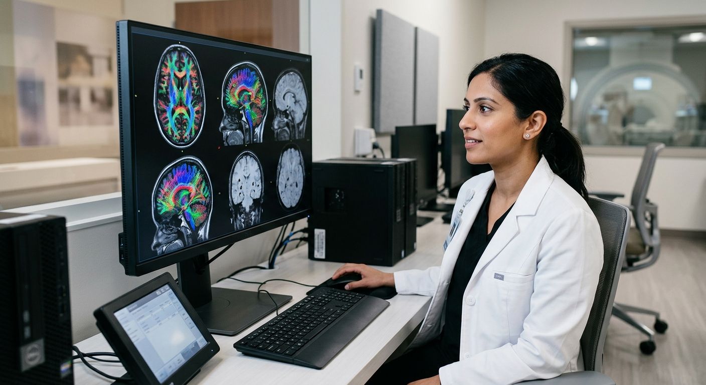
Board-certified radiologist reviewing advanced brain MRI scans on a large monitor in a modern imaging suite
Healthcare Legal Support, Medical Imaging Services, MRI & CT Diagnostics, Personal Injury Care, Radiology Protocols, Traumatic Brain Injury (TBI)

Traumatic Brain Injury Diagnostic Standards: DTI and SWI Insights

Table of Contents Understanding Traumatic Brain Injury Diagnostic Standards Clinical Frameworks and Guidelines for TBI Diagnosis Advanced MRI Techniques for TBI Detection Diffusion Tensor Imaging in Traumatic Axonal Injury Susceptibility Weighted Imaging and Microhemorrhage Detection Integrating Serum Biomarkers with Imaging Implementing TBI Diagnostic Imaging in Clinical and Legal Practice Coordinating with Physicians and Attorneys for Timely Imaging Streamlining Prior Authorizations and Liens for TBI Cases Ensuring Rapid Report Turnaround and Legal Documentation Biopsychosocial and Multimodal Approaches to TBI Assessment Frequently Asked Questions About TBI Diagnostic Standards Advancing TBI Care Through Accurate Diagnostic Imaging Understanding Traumatic Brain Injury Diagnostic Standards Understanding traumatic brain injury diagnostic standards is essential for accurate imaging and patient care. At Arizona Advanced Imaging LLC, we rely on proven protocols to detect both obvious and subtle brain injuries, guiding every step from initial assessment to comprehensive evaluation. Our board-certified neuroradiologists utilize standardized criteria to ensure clarity in every image and confidence in every decision. Computed tomography (CT) remains the first-line standard for acute TBI. Its speed in detecting hemorrhages and fractures makes it indispensable in emergency settings. For subacute and chronic evaluations, magnetic resonance imaging (MRI) offers superior resolution, revealing diffuse axonal injuries or small contusions that CT may miss. We adhere to these established standards because timely, accurate diagnosis directly shapes treatment planning and recovery outcomes. Emerging advanced techniques, such as diffusion tensor imaging and susceptibility weighted imaging, are refining traumatic brain injury diagnostic standards. DTI maps white matter tracts to detect microstructural damage, while SWI heightens sensitivity to minute hemorrhages. Our fellowship-trained specialists employ these modalities when standard imaging proves inconclusive, ensuring no injury remains hidden. Validated scoring systems and structured reporting further strengthen the medicolegal value of every study, supporting both clinical decisions and legal documentation. These standards evolve with injury severity and time since injury. For a broader overview, explore the patient guide to medical imaging. Please consult with a healthcare professional for diagnosis and treatment advice, as results may vary based on individual patient conditions and imaging quality. Clinical Frameworks and Guidelines for TBI Diagnosis Having examined the fundamentals of TBI diagnosis, we now compare the major clinical frameworks shaping current assessment. Current traumatic brain injury diagnostic standards draw from three complementary models, each informing the way we structure our TBI care imaging protocols. As Your Trusted Radiology Partner, we apply these evidence-based frameworks to every TBI evaluation. The 2026 AAN/CDC guidelines standardize diagnosis across care settings, relying on CT for acute triage and MRI for follow-up, with serum biomarkers S100B, GFAP, and UCH-L1 guiding imaging decisions. According to RadiologyInfo, a patient education resource from RSNA and ACR, CT remains first-line for acute head trauma assessment. The CBI-M framework classifies injury severity for targeted treatment using advanced MRI, including diffusion tensor imaging and susceptibility weighted imaging, to detect microstructural injuries like diffuse axonal injury that conventional scans frequently miss. Our Spin Tech MRI protocol employs these same advanced sequences, delivering quantitative DTI data and automated microbleed detection that directly support the CBI-M classification system and its treatment stratification approach. The four pillars of modern assessment integrate clinical examination, advanced imaging, biomarker analysis, and outcome tracking into a unified diagnostic approach. Beyond DTI and susceptibility weighted imaging, this model incorporates functional MRI for evaluating brain activity patterns following injury. The biomarker panel expands to include neurofilament light alongside GFAP and UCH-L1, enabling broader assessment of neuronal damage and recovery. Notably, the CBI-M framework does not explicitly define biomarker use, unlike both the 2026 guidelines and the four-pillar model. The following table provides a side-by-side comparison of these three diagnostic frameworks: Comparison of TBI Diagnostic Frameworks 2026 Clinical Guidelines CBI-M Framework Four Pillars of Modern Assessment Standardize diagnosis across care settings Classify injury severity for targeted treatment Integrate clinical, imaging, biomarker, and outcome data CT for acute triage, MRI for follow-up Advanced MRI (DTI/SWI) for microstructural injury DTI, SWI, functional MRI for comprehensive evaluation S100B, GFAP, UCH-L1 recommended Not explicitly defined GFAP, UCH-L1, neurofilament light integrated Advanced MRI Techniques for TBI Detection Diffusion Tensor Imaging in Traumatic Axonal Injury Diffusion tensor imaging measures the directional movement of water molecules along white matter tracts in the brain. In healthy tissue, water diffuses predictably along axonal bundles, but when axons are sheared or damaged, this organized diffusion becomes disrupted. The result is a measurable reduction in fractional anisotropy, a metric that reflects white matter integrity. As a fellowship-trained team, we at Arizona Advanced Imaging use quantitative DTI to detect these diffuse axonal injuries, which are often invisible on conventional MRI sequences. Our spin tech MRI TBI protocol incorporates DTI tractography to map white matter pathways and identify regions of reduced connectivity. Even in mild TBI cases where patients report persistent cognitive symptoms, susceptibility weighted imaging and diffusion tensor imaging can reveal objective evidence of injury that would otherwise go undetected. Susceptibility Weighted Imaging and Microhemorrhage Detection Susceptibility weighted imaging exploits magnetic susceptibility differences between tissues to detect blood products that accumulate after traumatic shearing forces tear small vessels. SWI is particularly effective at visualizing microhemorrhages and hemosiderin deposits that appear as tiny dark foci scattered throughout the brain parenchyma. Comparison of DTI and SWI for TBI Detection Comparison of DTI and SWI for TBI Detection Diffusion Tensor Imaging (DTI) Susceptibility Weighted Imaging (SWI) Measures water diffusion along white matter tracts Detects magnetic susceptibility differences from blood products Reduced fractional anisotropy indicating axonal injury Microhemorrhages, hemosiderin deposits High sensitivity for diffuse axonal injury Moderate sensitivity; better for hemorrhagic shear injuries Prognostic biomarker for cognitive outcome Identifies lesion burden correlating with severity DTI and SWI comparison for advanced TBI detection Integrating Serum Biomarkers with Imaging Emerging blood-based biomarkers offer an additional layer of diagnostic information that complements advanced MRI findings. After traumatic brain injury, proteins such as glial fibrillary acidic protein, ubiquitin C-terminal hydrolase L1, and neurofilament light are released from damaged neural cells into the bloodstream. Implementing TBI Diagnostic Imaging in Clinical and Legal Practice Coordinating with Physicians and Attorneys for Timely Imaging Successful TBI case

Scroll to Top