Open MRI vs Closed MRI Accuracy 2026: A Definitive Diagnostic Guide
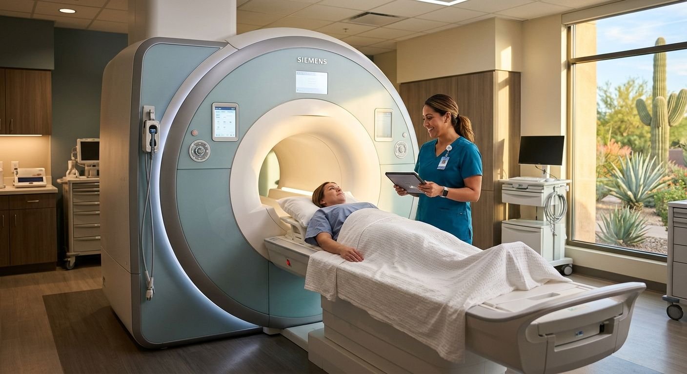
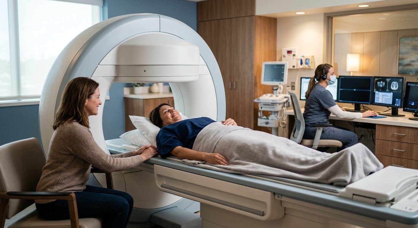
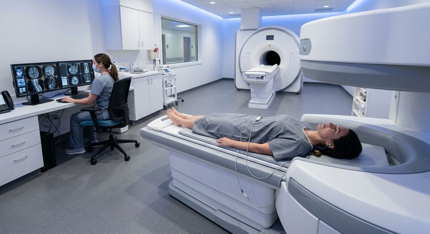
Table of Contents Demystifying MRI Accuracy: Open vs Closed Systems 1. MRI Accuracy: Open vs. Closed in 2026 2. Open MRI Solutions for Severe Claustrophobia 3. Closed MRI: The Gold Standard for High-Resolution Brain Scans 4. TBI Detection Accuracy with Open MRI Protocols 5. Scan Time Differences Between Open and Closed MRI 6. High-Field Open MRI: Bridging the Diagnostic Sensitivity Gap 7. Musculoskeletal Imaging: Open MRI Resolution Capabilities 8. Making Informed Decisions for Personal Injury Imaging Rapid, Reliable MRI Results for Your Legal and Medical Needs Demystifying MRI Accuracy: Open vs Closed Systems When evaluating open MRI vs closed MRI accuracy in 2026, a critical factor often overlooked is how patient anxiety influences scan quality. Traditional closed-bore units, with their narrow, tube-like design, can trigger intense claustrophobia, causing involuntary movement, or requiring sedation, which introduces motion artifacts that blur the final images. Open MRI technology uses two large flat magnets instead of a narrow tube, giving patients more space and reducing claustrophobic triggers. This comfort allows many patients to complete their exam without sedation, minimizing motion blur and preserving image integrity. At Arizona Advanced Imaging, we see how this reduces repeat scans and supports faster, clearer diagnoses for personal injury cases. Modern open MRI systems deliver high-resolution studies that support accurate clinical decisions across a wide range of conditions. 1. MRI Accuracy: Open vs. Closed in 2026 Open MRI systems use two large, flat magnets positioned above and below the patient, as opposed to the traditional long, narrow magnet. The design reduces the claustrophobic feelings associated with MRI. Open MRI eases claustrophobia; Closed MRI delivers highest spatial resolution. When patients feel anxious or move inside a closed MRI, even slight motion can create blurring and artifacts. Our team at Arizona Advanced Imaging finds that the open design minimizes this risk: when patients remain comfortable, motion artifacts are greatly reduced, often eliminating the need for sedation. While a traditional closed system gives higher resolution images due to a stronger magnetic field, a system free from artifacts is still diagnostically sufficient. As your trusted radiology partner, we recognize that patient comfort is critical, especially for claustrophobic patients who often cannot remain still. 2. Open MRI Solutions for Severe Claustrophobia Of course, we understand the importance of diagnosis. But for patients with debilitating claustrophobia, comfort and convenience also play a key role in getting clear images. Open MRI replaces the traditional enclosed MRI machine with a large, flat open design. This machine removes the confining tunnel sensation that triggers anxiety and lets patients see around the room, reducing feelings of restriction during the scan. In a conventional closed MRI, distress can cause involuntary movement and motion artifacts that compromise image quality and require repeat scans. The spacious design of our open system reduces anxiety so many claustrophobic patients complete their exams without sedation. Your health and clarity are our priority. We offer same-day or next-day appointments and provide reports within 24 to 48 hours to avoid delays. While a closed MRI in Phoenix remains the gold standard for certain high-resolution neurological studies, open MRI provides a viable alternative for patients who cannot tolerate an enclosed environment. 3. Closed MRI: The Gold Standard for High-Resolution Brain Scans According to our patient resources, open MRI offers a comfortable alternative for claustrophobic individuals, yet closed MRI remains the gold standard for high-resolution brain scans. Closed MRI systems use stronger magnetic fields, typically 1.5T or 3T, which deliver the fine anatomical detail necessary to detect subtle brain abnormalities. At our imaging centers across Arizona and Texas, we maintain both closed and open MRI systems so that referring providers can select the optimal modality for each patient’s diagnostic requirements. For brain imaging, our radiologists recommend closed MRI as the first-line choice when patient tolerance permits. Only if severe claustrophobia or anxiety would prevent a diagnostic-quality scan do we proceed with open MRI using adapted protocols. This high-field resolution is particularly critical for identifying subtle traumatic brain injury findings that may be missed on lower-strength open systems. With a standard 24- to 48-hour report turnaround, clinicians can act on results quickly to guide neurological treatment decisions. 4. TBI Detection Accuracy with Open MRI Protocols While closed MRI is the gold standard for brain imaging, at Arizona Advanced Imaging, our open MRI units are specialized to perform particular sequences (diffusion weighted and susceptibility weighted imaging) to more effectively identify brain injuries. These sequences help us reliably detect traumatic brain injury (TBI), visualizing subtle hemorrhages and axonal injuries that go undetected on routine brain images. A fellowship-trained neuroradiologist reviews the images, applying specialized expertise to identify and interpret findings that support an accurate diagnosis. This expert interpretation is as critical as the imaging itself, ensuring that our reports meet the standards required for clinical decisions and legal documentation. For patients who experience claustrophobia, our open MRI offers a comfortable alternative without compromising ability to run these TBI-specific sequences, ensuring that those who might otherwise avoid imaging receive the diagnostic evaluation they need. The tbi detection accuracy open mri achieves depends on the protocol design and the neuroradiologist’s expertise, not merely the scanner type. 5. Scan Time Differences Between Open and Closed MRI Having discussed how open MRI protocols aid in the diagnosis of TBI, we should consider how long each exam takes. It is actually more dependent on patient cooperation than MRI scanner type. In closed MRI, the confined tube often provokes claustrophobia. Anxious patients may shift involuntarily, creating motion artifacts that blur images and force technologists to rerun sequences. At Arizona Advanced Imaging, we see that open MRI’s spacious design—where the patient lies between two flat magnets—greatly reduces such distress, lowering the repeat rate. Even though a closed scanner’s higher field strength might theoretically shorten individual acquisitions, the effective exam time can be longer when factoring in rescanning. Other variables, such as coil design and the area being imaged, further affect duration, making simple minute-per-minute comparisons misleading. Various time-related factors highlight the broader effort in imaging to combine patient