

Table of Contents
- The SpinTech MRI TBI Protocol: A New Standard in Brain Injury Detection
- How the SpinTech Protocol Detects Micro-Hemorrhages and Axonal Injury
- Exploring Quantitative Susceptibility Mapping and Diffuse Axonal Injury Detection
- Accessing the SpinTech Protocol for TBI Imaging Across Arizona and Texas
- Frequently Asked Questions About the SpinTech MRI TBI Protocol
- Advancing TBI Diagnosis with the SpinTech MRI Protocol
The SpinTech MRI TBI Protocol: A New Standard in Brain Injury Detection
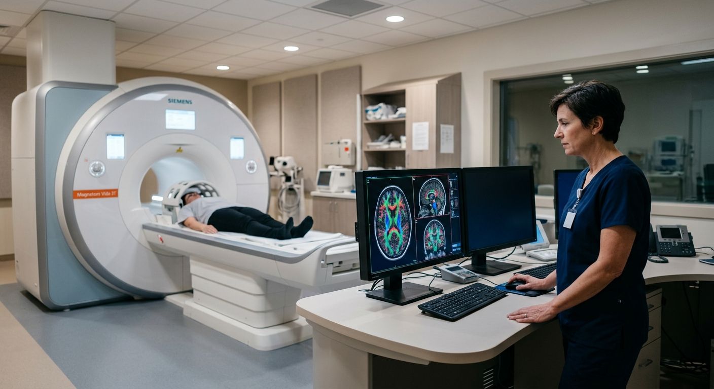
Conventional MRI plays a critical role in brain imaging, yet it can miss up to half of the subtle microbleeds associated with mild traumatic brain injury. We address this diagnostic gap with the SpinTech MRI TBI protocol, a proprietary advanced imaging method available at AZ Advanced Imaging. By moving beyond standard structural scans, this approach provides objective evidence of injury that standard imaging often overlooks, helping patients and their physicians make informed decisions.
This advanced TBI imaging protocol combines several specialized sequences to reveal what conventional scans cannot. Quantitative susceptibility mapping, or QSM, detects tiny deposits of iron from blood products, pinpointing microbleeds with exceptional sensitivity. Diffuse axonal injury detection is achieved through diffusion tensor imaging, or DTI, which assesses the integrity of white matter tracts and uncovers microstructural damage that disrupts normal brain communication. Susceptibility-weighted imaging, or SWI, further enhances the visualization of these otherwise invisible injuries. Every scan is interpreted by our board-certified, fellowship-trained radiologists, and we deliver a detailed report within 24 to 48 hours, ensuring you receive timely, actionable results.
Clarity in every image, confidence in every decision. Our advanced TBI imaging protocol is particularly suited for personal injury patients who need objective and defensible documentation of brain injury. Results may vary based on individual patient conditions. We serve patients throughout Arizona and Texas from our primary location in Phoenix, providing access to technology that can make a decisive difference in both clinical care and legal proceedings.
How the SpinTech Protocol Detects Micro-Hemorrhages and Axonal Injury
Our spintech mri tbi protocol addresses the critical limitations of standard imaging by applying advanced sequences specifically designed to identify the subtle markers of mild traumatic brain injury. Where conventional scans may fail to detect up to 50% of cerebral microbleeds, this quantitative approach provides objective, measurable, and defensible results essential for both clinical treatment and legal documentation.
Enhanced Detection of Micro-Hemorrhages
The first pillar of the protocol’s diagnostic power lies in Susceptibility Weighted Imaging (SWI) and its advanced post-processing technique, quantitative susceptibility mapping. SWI is an MRI sequence exquisitely sensitive to hemosiderin, a blood breakdown product that accumulates in the brain after a micro-hemorrhage. On SWI images, these tiny bleeds appear as small, dark foci—findings that are often invisible on conventional scans. Our fellowship-trained, board-certified neuroradiologists then utilize QSM to go a step further, calculating the actual magnetic susceptibility of these lesions. This quantification allows for a crucial differentiation between acute and chronic micro-hemorrhages, mapping the full extent and age of the injury with a precision standard imaging cannot achieve.
Identifying Diffuse Axonal Injury
A second cornerstone of the SpinTech approach is diffuse axonal injury detection through Diffusion Tensor Imaging (DTI). While conventional MRI may show moderate sensitivity to this type of trauma, DTI provides a microscopic view of the brain’s white matter architecture. It works by measuring the directional diffusion of water molecules, which normally flow freely along intact axonal fiber tracts. When a shearing injury occurs—a hallmark of TBI from car accidents or falls—this orderly flow is disrupted. The protocol quantifies this disruption as a reduction in fractional anisotropy, a metric that serves as a direct, evidence-based indicator of axonal injury. This capability makes our protocol highly sensitive for detecting diffuse axonal injury that may otherwise go undiagnosed.
The table below highlights key differences between a conventional brain MRI and our FDA-approved SpinTech protocol.
| Feature | Conventional MRI | SpinTech MRI TBI Protocol |
|---|---|---|
| Ability to detect micro-hemorrhages | Limited | High sensitivity with SWI and QSM |
| Scan time for brain | 20-30 minutes | 10-15 minutes (up to 50% faster) |
| Sensitivity to diffuse axonal injury | Moderate | High with DTI and SWI |
| Requirement for contrast agent | Often needed | Typically non-contrast |
By eliminating the need for a contrast agent in most cases and completing the scan in as little as 10 to 15 minutes, this non-contrast protocol significantly reduces patient risk and accelerates the diagnostic process. This combination of speed, safety, and sensitivity provides a clear advantage in building a comprehensive clinical picture.
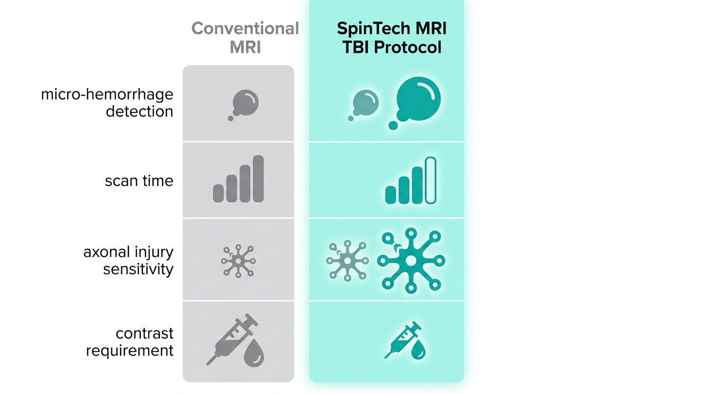
SpinTech MRI TBI Protocol vs. Conventional MRI comparison
This integrated multi-sequence strategy is the foundation of our TBI diagnostic imaging legal support. By providing court-ready reports that objectively document the presence and nature of an injury, we give personal injury attorneys and physicians the clarity they need to make confident decisions for their cases and patients.
Exploring Quantitative Susceptibility Mapping and Diffuse Axonal Injury Detection
The Role of Quantitative Susceptibility Mapping (QSM)
The SpinTech MRI TBI protocol represents an advanced imaging strategy that we employ to enhance the detection of subtle traumatic brain injuries. Building on the fundamental sensitivity of susceptibility weighted imaging, we now examine how quantitative susceptibility mapping (QSM) refines the identification of diffuse axonal injury. QSM is a post-processing technique that quantifies magnetic susceptibility within brain tissues, generating high-resolution maps that differentiate diamagnetic calcifications from paramagnetic blood products such as micro-hemorrhages.
Detecting Diffuse Axonal Injury with Advanced Sequences
Diffuse axonal injury detection relies on a multi-sequence approach that evaluates white matter integrity and micro-hemorrhage burden concurrently. Diffusion tensor imaging (DTI) is the primary tool for assessing the structural connectivity of white matter tracts, generating scalar metrics such as fractional anisotropy that decline when axons are sheared during acceleration-deceleration forces.
| Technique | Sensitivity to Micro-hemorrhages | Specificity for DAI | Clinical Utility |
|---|---|---|---|
| Susceptibility Weighted Imaging (SWI) | Very high | Moderate | Widely used, qualitative |
| Quantitative Susceptibility Mapping (QSM) | High | High | Quantitative, advanced |
| Conventional Gradient Echo (GRE) | Moderate | Low | Standard, less sensitive |
Accessing the SpinTech Protocol for TBI Imaging Across Arizona and Texas
For patients and legal professionals ready to access the spintech mri tbi protocol, we offer convenient imaging locations across Arizona and Texas. This advanced imaging technology provides objective, court-ready documentation of traumatic brain injuries that standard scans often miss.
Finding Imaging Centers Equipped with the SpinTech Protocol
Our eight Arizona locations serve communities including Phoenix, Scottsdale, Mesa, Tucson, Glendale, Chandler, Gilbert, and Tempe. In Texas, multiple centers provide coverage across Dallas, Houston, San Antonio, Austin, and Fort Worth. Each facility is equipped with advanced MRI technology capable of running the SpinTech protocol.
What to Expect: Referral, Authorization, and Report Turnaround
The process begins when your referring physician or attorney contacts our scheduling team. We handle insurance verification and pre-authorization on your behalf. Most reports are ready within 24 to 48 hours and delivered digitally to your referring physician and legal team.
Coordinating with Legal Teams for Court-Ready Imaging
When TBI imaging is intended for litigation, we follow strict chain-of-custody procedures from the moment your scan begins. All imaging data is securely stored and provided in a format admissible in court. The spintech mri tbi protocol is particularly valuable in legal contexts because it enables diffuse axonal injury detection with objective precision.
Frequently Asked Questions About the SpinTech MRI TBI Protocol
What is the SpinTech MRI TBI protocol?
The SpinTech MRI TBI protocol is a specialized, non-invasive MRI examination designed specifically for assessing traumatic brain injury. It uses advanced sequences including susceptibility-weighted imaging (SWI), diffusion tensor imaging (DTI), and quantitative susceptibility mapping (QSM) to detect subtle brain changes often missed by standard scans.
Advancing TBI Diagnosis with the SpinTech MRI Protocol
At Arizona Advanced Imaging, we employ the SpinTech MRI protocol—an advanced imaging tool that combines susceptibility-weighted imaging (SWI), quantitative susceptibility mapping (QSM), and diffusion tensor imaging (DTI) to detect traumatic brain injury biomarkers often invisible on standard scans. Conventional MRI can miss up to 50% of cerebral microbleeds, making this protocol essential for accurate diagnosis and documentation.
Questions about imaging or scheduling? Call 844-641-2111 or find a location to contact your preferred imaging center.