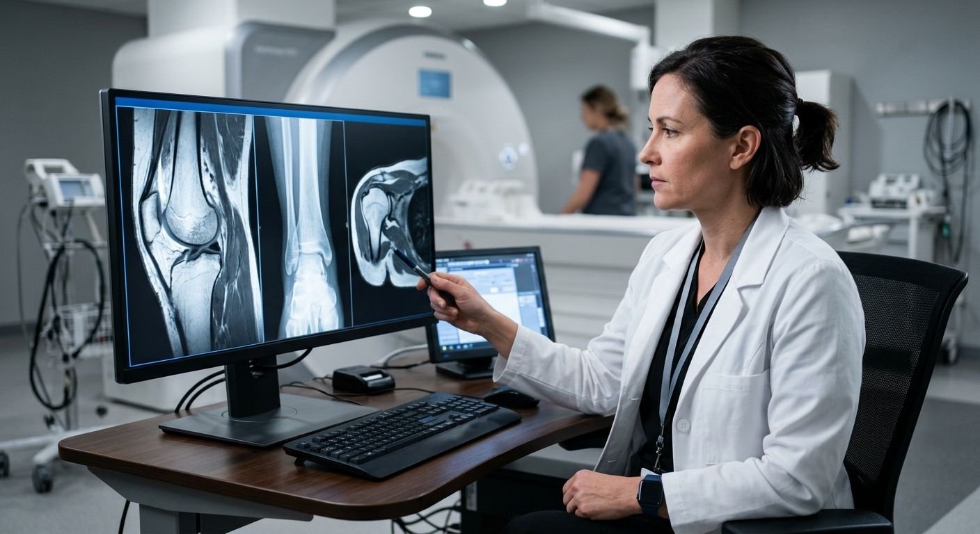
Imaging Tests for Arthritis Diagnosis: A Complete Medical Guide
Table of Contents How Medical Imaging Brings Clarity to Arthritis Diagnosis Imaging Tests for Arthritis Diagnosis: Common Modalities and Their Roles MRI vs. X-Ray: Deciding on Imaging for Joint Pain Ultrasound's Contribution to Diagnosing Inflammatory Arthritis Distinguishing Osteoarthritis from Inflammatory Arthritis Through Imaging Preparing for Your Arthritis Imaging Appointment and Understanding Your Report Moving Forward: Getting the Right Imaging for Your Arthritis Your Next Steps Toward a Definitive Arthritis Diagnosis How Medical Imaging Brings Clarity to Arthritis Diagnosis Imaging tests for arthritis diagnosis provide the objective evidence needed to identify joint damage and inflammation that physical examination alone may miss. Arthritis symptoms, pain, swelling, stiffness, often overlap with other conditions, making an accurate diagnosis difficult without visual confirmation. At Arizona Advanced Imaging, we address this challenge with a comprehensive range of modalities including MRI, CT, X-ray, and ultrasound. Our board-certified, fellowship-trained radiologists in musculoskeletal imaging interpret these studies, revealing subtle signs of rheumatoid arthritis, osteoarthritis, and other joint diseases. Our imaging centers adhere to American College of Radiology safety and quality standards, ensuring high-resolution images and reliable reports delivered within 24 to 48 hours. Clarity in every image, confidence in every decision. While imaging supports accurate diagnosis, it complements, not replaces, a thorough clinical evaluation. From X-rays highlighting bone erosion to MRI’s detailed views of cartilage and soft tissue, different modalities each contribute unique insights that guide effective treatment planning. Key Takeaways Imaging Tests for Arthritis Diagnosis: Common Modalities and Their Roles Getting an accurate diagnosis for arthritis usually requires imaging tests which can show what your bones and soft tissue look like inside your body. As your trusted radiology partner, we assist our patients and their doctors in finding the right choice. Imaging Modality Best For Key Advantages Limitations Typical Role in Arthritis Diagnosis X-Ray Joint space narrowing, osteophytes, bone erosions Quick, low radiation, widely available Limited soft-tissue detail; cannot detect early cartilage loss First-line for osteoarthritis, screening for joint damage MRI Soft tissues: cartilage, synovium, bone marrow No ionizing radiation, excellent soft-tissue contrast, detects early inflammation Longer scan time, higher cost, may require contrast Problem-solving for inflammatory arthritis, detecting bone marrow edema CT Scan High-resolution bone detail, complex fractures Rapid acquisition, 3D reconstructions, superior bone visualization Higher radiation dose, limited soft-tissue contrast Preoperative planning, complement to MRI for bony anatomy Ultrasound Dynamic synovitis, fluid collections, tendon movement No radiation, real-time, inexpensive, can guide injections Operator-dependent, limited penetration depth First-line for peripheral joint inflammation, guiding aspirations Comparison of Imaging Modalities for Arthritis Diagnosis The x-ray is usually the most used initial imaging test because it can show the bone structure, joint space narrowing, osteophytes, and erosion. It is also useful for confirming osteoarthritis and for monitoring disease progression. Magnetic resonance imaging (MRI) is great for looking at soft tissue areas including cartilage, joint lining (synovium), and bone marrow. It can help see signs of inflammation that may appear in bone marrow ahead of actual tissue damage. MRI uses no ionizing radiation. CT provides high-resolution cross-sectional bone images that reveal subtle fractures and complex anatomy not seen on X-ray. Its 3D reconstructions aid in surgical planning. CT is reserved for cases where detailed bone assessment is crucial. Using ultrasound imaging, which employs sound waves to image soft tissues, is excellent for assessing synovial thickening, fluid, and tendon movement; it is radiation-free, can guide injections, and is a first-line tool for peripheral joint inflammation. The choice of modality depends on whether osteoarthritis or an inflammatory arthritis is suspected. With a clear understanding of each, we can now consider how MRI and X-ray compare when evaluating joint pain. MRI vs. X-Ray: Deciding on Imaging for Joint Pain When joint pain restricts mobility, diagnostic imaging becomes a key tool for uncovering the source. X-rays and MRIs are two types of images that may be taken during an evaluation. Knowing what these tests can reveal will help you better understand your diagnostic pathway if your healthcare professional recommends one test over another. An x-ray image captures bone structure using only a small dose of ionizing radiation. This type of test can be used to detect any fractures or dislocations in bones, as well as any bone spur growths and reduced joint spaces that are associated with osteoarthritis. As a quick procedure that is also easy to perform digitally, an x-ray will usually be the first form of imaging requested when joint pain is evaluated. MRI works without radiation, uses powerful magnets and radio waves to produce high-resolution images of soft tissues. It is especially good for showing cartilage, tendons, ligaments, muscles, and bone marrow, often revealing inflammation or early degenerative changes. Therefore, if a ligament tear, meniscus injury, or inflammatory condition such as rheumatoid arthritis is suspected, or when an X-ray result does not explain the patient’s symptoms, a doctor may order an MRI to get more information. At Arizona Advanced Imaging, we offer both digital X-ray (available on a walk-in basis) and MRI in open and closed configurations for patients experiencing joint pain in Arizona and Texas. Our open MRI systems help individuals with claustrophobia undergo a comfortable scan while maintaining diagnostic image quality. X-ray vs. MRI: complementary imaging for joint pain diagnosis. Whether to order an x-ray, MRI or both will be determined by the referring physician based on the patient’s history, physical exam findings, and what structures need to be evaluated. Frequently they may be used together to give the physician more information for diagnosis and treatment. Beyond X-ray and MRI, other imaging tools such as ultrasound also contribute to arthritis evaluation by detecting joint inflammation and guiding therapeutic injections. Your health and clarity are our priority, and we work closely with referring providers to coordinate the right imaging plan. Please consult your physician to determine which test is appropriate for your situation. Ultrasound’s Contribution to Diagnosing Inflammatory Arthritis When considering imaging tests for arthritis diagnosis, ultrasound provides a dynamic and non-invasive method for detecting early signs of inflammatory arthritis, such as synovitis and joint effusion. Unlike X-ray, which primarily