

Table of Contents
- Dynamic MSK Ultrasound Benefits: Real-Time Solutions for Soft Tissue Injuries
- The Technology Behind Dynamic Musculoskeletal Ultrasound
- Proven Benefits of Real-Time Musculoskeletal Ultrasound
- From Preparation to Report: The Dynamic MSK Ultrasound Procedure
- Integrating Dynamic MSK Ultrasound into Clinical and Legal Workflows
- Embracing Dynamic MSK Ultrasound for Comprehensive Musculoskeletal Care
Dynamic MSK Ultrasound Benefits: Real-Time Solutions for Soft Tissue Injuries
Dynamic musculoskeletal ultrasound provides clinicians with a window into soft tissue function that static imaging simply cannot offer. By capturing real-time movement of tendons, ligaments, muscles, and nerves during active motion or weight-bearing, this modality reveals biomechanical relationships that are invisible on a still image.
Unlike conventional MRI or X-ray that show anatomy at rest, dynamic ultrasound allows our team to assess how structures behave under stress. We can directly visualize a tendon gliding through a sheath, a nerve snapping over a bony prominence, or a ligament’s integrity during a pivot test. This makes it especially valuable for diagnosing tendon tears, nerve entrapment syndromes such as carpal tunnel, and joint instability in the shoulder, ankle, or wrist. The real-time feedback provides clarity in every image, confidence in every decision, critical for accurate diagnosis and personalized treatment planning.
The precise evaluation possible with dynamic ultrasound depends on advanced transducer technology, high-frequency imaging, and the expertise of a sonographer who can simultaneously position the patient, apply stress, and optimize the image. These technical foundations make real-time soft tissue assessment both practical and accessible for patients across Arizona and Texas.
The Technology Behind Dynamic Musculoskeletal Ultrasound
The dynamic musculoskeletal ultrasound is the foundation of the technology discussed in this paper. This technique captures real-time images of moving soft-tissue structures, revealing dysfunction that might otherwise remain hidden.
What Is Dynamic Musculoskeletal Ultrasound?
Dynamic musculoskeletal ultrasound uses high-frequency sound waves to visualize muscles, tendons, ligaments, nerves, and joints in real time. While MRI and X-ray produce excellent static images of structural anatomy, they cannot capture how tissues behave during movement or under load. With dynamic ultrasound, we apply a transducer to the skin and observe the area as the patient actively or passively moves. The system processes returning echoes into live video, enabling immediate assessment. It is particularly valuable when the source of pain emerges only during specific postures or activities. Our board-certified radiologists rely on high-resolution, real-time visualization to evaluate tendon gliding, ligament stability, and joint mechanics with a precision that static images alone cannot offer. This approach bridges the gap between static anatomy and functional pathology, providing critical diagnostic clues that complement other imaging modalities.
The Role of Real-Time Functional Assessment
The defining strength of dynamic musculoskeletal ultrasound is its capacity for real-time functional assessment. By continuously scanning while the patient performs active or passive maneuvers, we directly evaluate how joints, tendons, and soft tissues respond to physiological stress. For example, we can observe a rotator cuff tendon as the arm is raised, noting any impingement or abnormal excursion. This functional perspective goes beyond anatomy to reveal the biomechanics underlying symptoms. Our technologists work with patients to replicate provocative movements under controlled conditions, capturing the exact moment of dysfunction. This live visual feedback sharpens diagnostic accuracy and aids in planning interventions such as ultrasound-guided injections or physical therapy protocols. Unlike static images that present a single moment, dynamic ultrasound reveals the full range of motion and the interplay between muscles, tendons, and bony landmarks. The result is a more comprehensive picture that directly addresses the functional questions clinicians face.
Clinical Scenarios Where Dynamic Imaging Excels
Shoulder impingement: By scanning the subacromial space as the patient elevates the arm, we can see soft-tissue impingement that often correlates with their pain. Snapping hip syndrome: The transducer follows the iliotibial band or iliopsoas tendon, and isolates the structure responsible for the snap over the greater trochanter or pelvic brim. Ankle instability: Stress and talar tilt tests under ultrasound visualization allow us to assess laxity of the ligaments that standard weight-bearing X-rays cannot measure. These observations often guide patients away from unnecessary surgery and toward earlier, more appropriate intervention. In each of these instances, dynamic imaging provides evidence for clinical suspicion. This allows the referring physician to make an informed decision about treatment. The benefits of this technology—shorter wait times for diagnosis, directed treatment, and improved patient outcomes— stem from the ability to visualize pathology in motion. As your Trusted Radiology Partner, we use our expertise in dynamic musculoskeletal ultrasound to provide these benefits to patients daily.
Proven Benefits of Real-Time Musculoskeletal Ultrasound
Real-time musculoskeletal ultrasound, or dynamic MSK ultrasound, provides a live, moving view of soft tissues that static imaging cannot match. At Arizona Advanced Imaging, we apply it to deliver more accurate assessments for patients throughout the Phoenix metropolitan area.
Advantages Over Static MRI and CT Imaging
Unlike MRI and CT, which capture anatomy in a fixed position, dynamic ultrasound lets us evaluate soft tissues while the joint moves. This real-time capability is invaluable for detecting conditions that appear only during specific activities, such as snapping tendons or dynamic ligament instability. The exam is contrast-free and radiation-free. The American College of Radiology recognizes musculoskeletal ultrasound as a first-line tool for many evaluations. Comparing both sides under identical stress strengthens diagnostic confidence and reduces the likelihood of missed findings when static studies are inconclusive.
Real-Time Diagnosis of Shoulder Impingement
Shoulder impingement often involves transient contact between the acromion and rotator cuff that static MRI cannot replicate. Real-time ultrasound directly depicts acromiohumeral dynamics as the patient moves, pinpointing the phase when impingement occurs. Peer-reviewed work in the Journal of Ultrasound supports that objective visualization during motion improves diagnostic accuracy. For personal injury and clinical cases, documenting functional impairment in real time provides clear evidence for referring clinicians and legal teams, helping avoid repeat imaging and enabling earlier targeted care.
Uncovering Movement-Dependent Soft Tissue Tears
Some rotator cuff and ligament tears become apparent only when the tissue is stressed. Dynamic ultrasound reveals these motion-dependent injuries by scanning while the patient reproduces the painful movement, capturing gaps or abnormal friction that static images miss. Dynamic elastography studies further show that associated changes in tissue stiffness can increase sensitivity beyond static techniques. By documenting tears during the provoking maneuver, we give surgeons and therapists precise functional information to guide planning and rehabilitation.
Cost-Effective Imaging for Personal Injury Cases
For personal injury patients, minimizing redundant imaging is essential. Real-time ultrasound delivers diagnostic information in a single session, reducing the need for follow-up MRI or CT. The exam requires no contrast or radiation. Our practice accepts liens and letters of protection, so imaging does not impose upfront costs for qualifying personal injury cases. Immediate results support faster medical and legal decision-making without unnecessary delays.
Precision Guidance for Regenerative Medicine Injections
Real-time ultrasound provides live needle visualization for regenerative injections, allowing clinicians to guide the injectate precisely into the target while avoiding nerves and vessels. Evidence in ultrasound literature supports that real-time guidance improves placement accuracy and reduces misplacement risk. The ability to adjust needle trajectory in complex anatomy—such as the shoulder or hip—enhances procedural safety and treatment intent. We provide dynamic ultrasound technology to ensure these procedures are performed with precise visualization.
Real-time ultrasound is fundamental to our practice, delivering faster, more functionally relevant diagnoses and supporting clearer clinical and legal documentation. Our exam protocols build on these advantages to capture the essential images for every injury.
From Preparation to Report: The Dynamic MSK Ultrasound Procedure
A dynamic MSK ultrasound is a focused, efficient imaging experience designed to provide real-time insights into soft tissue function. Unlike static imaging methods, this exam involves active patient participation to capture how tendons, ligaments, and muscles move during specific actions.
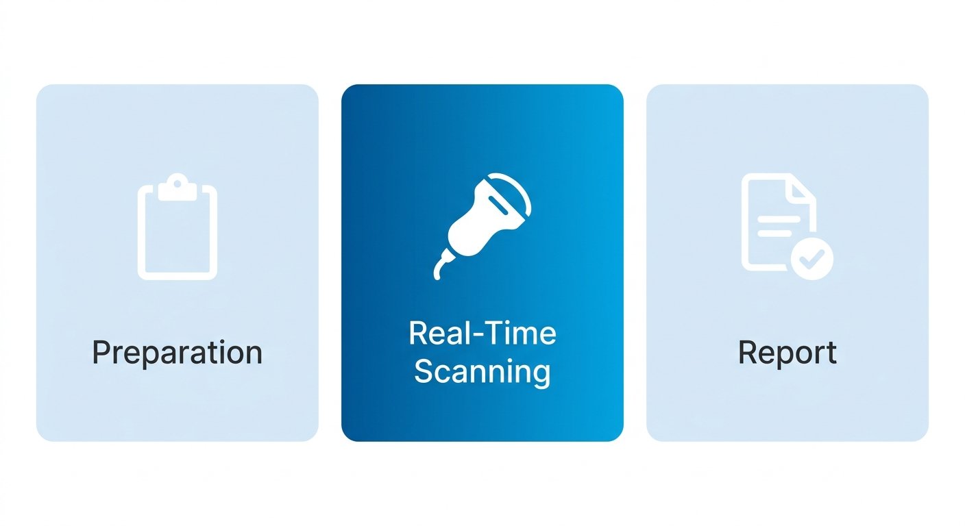
Knowing what to expect at each phase helps you arrive prepared and leave with confidence in your results.
Preparing for Your Dynamic MSK Ultrasound
Preparation for your dynamic MSK ultrasound begins with a few straightforward administrative steps. When you schedule your appointment, we coordinate directly with your referring physician to obtain the necessary referral documentation. Our team verifies your insurance benefits and handles pre-authorization requirements before your visit. For patients involved in personal injury cases, we accept letters of protection and lien paperwork, meaning there is no upfront cost required for imaging services. We also support workers’ compensation and third-party liability billing for active-duty military members, veterans, and TRICARE beneficiaries without requiring payment at the time of service.
On the day of your exam, physical preparation is simple. Wear loose, comfortable clothing that allows easy access to the joint area being examined. No fasting or sedation is needed. If you are wearing a cast, brace, or bandage, the technologist will work around it as needed to obtain clear images without compromising your comfort or care.
During the Examination: Real-Time Scanning and Patient Movements
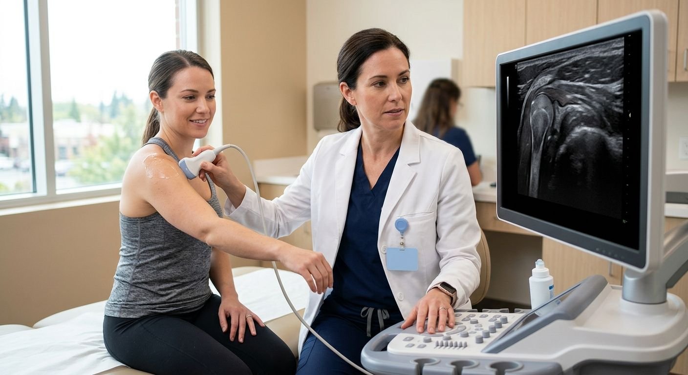
The technologist guides you through specific movements—bending a joint, lifting an arm, rotating a shoulder, or walking a few steps—while the radiologist observes your muscles, tendons, and ligaments in motion on a live screen. Unlike a static MRI where you must remain completely still, a dynamic ultrasound relies on your active participation.
Movement-based imaging captures how tissues behave only in positions that reproduce your symptoms, revealing abnormalities that might remain hidden in a stationary scan. The radiologist freezes key frames at the precise moment of discomfort or restricted motion, creating a functional record of the problem.
After the Scan: Rapid Interpretation and Report Availability
The captured images are forwarded to a board-certified radiologist with fellowship training in musculoskeletal imaging, who performs a detailed interpretation of the study. Our commitment to rapid turnaround means the finalized report is available to your referring physician or attorney within 24 to 48 hours.
They are structured as case-ready documentation that can substantiate injury claims, including evidence of pain and suffering, without requiring additional testimony or reformatting. We route the report securely to all authorized parties—physicians, case managers, and attorneys—through our provider portal and direct communication channels.
Integrating Dynamic MSK Ultrasound into Clinical and Legal Workflows
Dynamic MSK ultrasound offers a functional perspective that enhances both clinical diagnosis and legal documentation, fitting naturally into the workflows of personal injury attorneys, referring physicians, and patients. The following guidance explains how each group can integrate this exam to achieve more precise and actionable results.
For Personal Injury Attorneys: Securing Court-Ready Imaging
When a requesting attorney partners with a personal injury imaging center, such as ours in Phoenix, the process to obtain court ready documentation is simplified. Typically, the attorney will request a lien-based imaging order—often called a letter of protection—so the client can undergo the exam without upfront payment. We will then verify imaging coverage and work directly with the attorney’s office to streamline the process. During the imaging session, we capture dynamic stress views of the targeted anatomy and produce a forensic-quality report that includes a radiologist’s interpretation, delivering functional evidence beyond static images. To support litigation deadlines, we work to schedule the examination to coordinate with the independent medical exams and discovery deadlines to ensure reports and records are available to the attorney when needed.
For Referring Physicians: Maximizing Diagnostic Value
Dynamic MSK Ultrasound allows referring physicians to evaluate conditions that may only be exhibited with active movement. Tendinopathy and partial tendon tears as well as nerve entrapment and joint instability conditions may be better diagnosed with dynamic MSK US. The dynamic nature of the evaluation helps to elucidate the source of patient’s symptoms and guide the type and course of treatment. Since the evaluation is performed and interpreted by a radiologist, the information is structured to facilitate next steps in patient management.
For Patients: Ensuring a Smooth Imaging Experience
We ask that patients bring a valid photo ID, their insurance card, and a list of current medications, and we recommend loose, comfortable clothing; we verify coverage and explain any potential out-of-pocket responsibility before the exam, and patients will receive any specific instructions, such as hydration or medication adjustments, at the time of scheduling. For personal injury cases, we work with the law firm to make arrangements so the patient is not required to pay out of pocket. We also provide free transportation and multilingual services within our area. All exams are supervised by board-certified radiologists, so patients can feel confident in both their comfort and the accuracy of their results. All exams will be conducted under the ACR (American College of Radiology) safety and quality standards and protocols.
Embracing Dynamic MSK Ultrasound for Comprehensive Musculoskeletal Care
Dynamic MSK ultrasound provides a motion-sensitive element that static MRI or CT cannot capture. By incorporating this modality with our high-resolution cross-sectional imaging, we bring greater clarity to conditions such as tendon subluxation, dynamic impingement, and guided injection placement, all within a single patient encounter. MSK dynamic ultrasound’s ability to image structures during active movement and to compare contralateral anatomy improves detection of position-dependent abnormalities.
Together, these elements improve diagnostic confidence for the clinician and the documentation for the case. For patients, it often lessens the need for additional studies and provides clarity for their musculoskeletal condition. We integrate dynamic ultrasound into our comprehensive imaging workflow to ensure movement-dependent findings are never left unseen.
Imagine how a fully integrated MSK imaging approach can support more informed care decisions for you and your patients. Our team coordinates closely with referring clinicians and legal partners, and coordinates same-day and next-day appointments. Within 24 to 48 hours, you will have a physician-interpreted report, so you can move forward with the answers you need. Please reach out for anything you need.
Discover Dynamic MSK Ultrasound
Now that you have explored the capabilities of dynamic musculoskeletal ultrasound, consider taking the next step toward comprehensive diagnostic care. Arizona Advanced Imaging offers state-of-the-art ultrasound exams that deliver real-time functional assessment directly into your clinical or legal workflow. Their board-certified radiologists interpret each scan, providing clear, case-ready reports within 24 to 48 hours. With same-day appointments available, you can avoid delays and obtain the imaging evidence you need. Whether you are a physician, attorney, or patient, the team handles everything from insurance verification to lien-based coordination. We describe how these exams support diagnostic goals.