

Table of Contents
- A Closer Look at Imaging Tests for Gallbladder Problems
- Diagnostic Imaging Exams for Gallbladder Health
- Why Ultrasound Is the First-Line Imaging Test for Gallbladder Issues
- How CT and MRI Provide Detailed Gallbladder and Bile Duct Images
- How to Prepare for Your Gallbladder Imaging Appointment
- Interpreting Gallbladder Imaging Results and Next Steps
- Advancing Your Gallbladder Health with Clear Imaging
A Closer Look at Imaging Tests for Gallbladder Problems
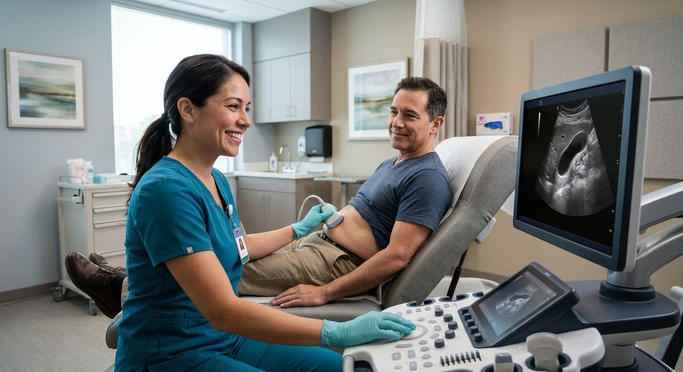
Imaging tests for gallbladder problems typically begin with an abdominal ultrasound, a noninvasive, radiation-free exam that uses sound waves to produce real-time images of the gallbladder. Because it involves no ionizing radiation, ultrasound is a safe first step for patients of any age and can be repeated when monitoring is needed. The exam can detect gallstones, wall thickening, and signs of cholecystitis, giving clinicians objective information to evaluate symptoms. At Arizona Advanced Imaging, we perform this study with certified sonographers and oversight by board-certified radiologists to ensure diagnostic accuracy. Our reports are structured for objective findings and can serve as legal documentation — a valuable benefit in personal injury cases where gallbladder symptoms are part of a claim. With same-day or next-day appointments and a typical 24- to 48-hour turnaround, we help you move from imaging to answers.
Here is a look at the various diagnostic tools used in examining the gallbladder.
Key Takeaways
- Evaluating gallbladder health relies on diagnostic imaging exams that assess both structure and bile flow.
- Among the diagnostic imaging options used for gallbladder concerns, ultrasound has been established as the first-line test by current clinical guidelines.
- When a gallbladder ultrasound detects an abnormality, advanced CT and MRI imaging provide the high-resolution, cross-sectional detail needed to characterize the finding and plan appropriate care.
- When the doctor prescribes gallbladder scanning, there are many things we need to pay attention to before performing imaging to help us obtain better quality diagnostic pictures.
- After your gallbladder scan has been completed, the radiologist will review the images taken and write up a description of what was seen on each image.
Diagnostic Imaging Exams for Gallbladder Health
Evaluating gallbladder health relies on diagnostic imaging exams that assess both structure and bile flow. Gallbladder disease affects about 8% of men and 17% of women, making accurate imaging essential. Ultrasound is the primary exam for suspected gallbladder disease, offering high sensitivity and specificity for detecting gallstones. According to the American College of Emergency Physicians, emergency physicians achieve sensitivity exceeding 86% and specificity over 88% when using ultrasound to identify gallstones. This noninvasive test uses high-frequency sound waves to create real-time images of the gallbladder and bile ducts, showing gallstones, wall thickening (normal ≤3 mm), duct dilation, and pericholecystic fluid. The exam also evaluates for a sonographic Murphy sign—tenderness when the probe presses over the gallbladder—which, combined with gallstones, yields a positive predictive value of 92% for acute cholecystitis.
When structural imaging alone cannot explain symptoms, a hepatobiliary iminodiacetic acid (HIDA) scan provides a functional assessment of bile flow. A radioactive tracer is injected into a vein and travels through the liver, gallbladder, and small intestine. Patients must fast for four to six hours; the initial scan takes up to 60 minutes. Nursing mothers should pause breastfeeding for one to two days after the procedure because the tracer passes into breast milk, as advised by Alberta Health Services.
HIDA scans are often recommended when ultrasound findings are inconclusive, as they provide functional detail that structural imaging cannot. While ultrasound excels at showing gallstones and anatomical changes, the HIDA scan reveals how well the gallbladder contracts and empties.
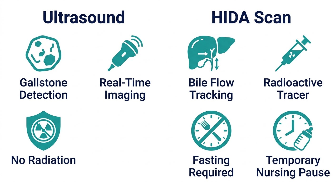
Together, these exams provide complementary information. Ultrasound’s role as a first-line test stems from its diagnostic accuracy, safety, and widespread availability. As your trusted radiology partner, Arizona Advanced Imaging offers abdominal ultrasound for gallbladder evaluation at our centers across Arizona and Texas. With over 25 years of experience in diagnostic imaging, we deliver consistent quality and compassionate patient care. Our licensed technologists and board-certified radiologists work together to provide accurate, timely imaging that supports your clinical care.
Why Ultrasound Is the First-Line Imaging Test for Gallbladder Issues
Among the diagnostic imaging options used for gallbladder concerns, ultrasound has been established as the first-line test by current clinical guidelines. The American College of Emergency Physicians (ACEP) identifies ultrasound as the preferred initial imaging modality for suspected biliary tract disease, a condition that ranks as the third most common cause of acute abdominal pain in emergency settings.
One key advantage of ultrasound is that it relies on high-frequency sound waves rather than ionizing radiation. This makes it safe for repeated examinations and for patients who cannot undergo CT scans. Ultrasound also provides real-time imaging, which allows physicians to assess gallbladder wall thickness, detect gallstones, and evaluate the common bile duct dynamically. ACEP data indicate that emergency physicians achieve sensitivity greater than 86% and specificity above 88% for identifying gallstones with point-of-care ultrasound. Furthermore, the combination of gallstones and a positive sonographic Murphy’s sign yields a 92% positive predictive value for cholecystitis. These strong performance characteristics, combined with the non-invasive nature and immediate availability of ultrasound, reinforce its role as the optimal starting point for gallbladder imaging.
We have several imaging facilities in Arizona and Texas that can perform abdominal ultrasounds. Each study is performed by a certified sonographer who follows the standardized protocols we follow for each patient. All examinations are interpreted by our board-certified radiologists who specialize in abdominal imaging. This clinical oversight ensures accurate, actionable reports without unnecessary delays, supporting both diagnostic clarity and timely treatment decisions.
Ultrasound is usually sufficient for initially diagnosing problems in the gallbladder, but more complex or inconclusive cases may require complementary imaging. In such cases, if the image obtained through ultrasound is of insufficient clarity because the ultrasound is too difficult for the patient’s body shape or weight, or when a detailed anatomical assessment of the bile ducts is needed, a CT scan or magnetic resonance imaging can provide additional information. Some advanced imaging may follow a routine ultrasound to provide additional detail.
How CT and MRI Provide Detailed Gallbladder and Bile Duct Images
When a gallbladder ultrasound detects an abnormality, advanced CT and MRI imaging provide the high-resolution, cross-sectional detail needed to characterize the finding and plan appropriate care. Ultrasound remains the preferred initial test for gallbladder symptoms, as guidance from the American College of Emergency Physicians underscores, but its ability to evaluate deep tissue extension or distant spread is limited. CT and MRI fill that gap, offering the precision required for cancer staging and treatment decisions.
CT scans produce pictures from numerous thin, detailed cross-sectional slices of tissue that show the size and shape of a gall bladder tumour, its invasion into nearby structures, and any spread to the liver or lymph nodes. Contrast material injected during the scan highlights blood vessels and abnormal tissue, making it easier to define the tumour’s borders. MRI scans can be done using strong magnetic fields and radio waves without requiring ionizing radiation. They provide exceptional soft-tissue contrast and they are highly effective for looking at the bile ducts since an additional special scan technique, MRCP, which stands for Magnetic Resonance Cholangiopancreatography, gives highly detailed and non-invasive images of the whole bile duct system. According to Cancer Research UK, imaging procedures like these play a major role in deciding how big the cancer is, how much it has spread through the liver and how it may have affected the bile ducts prior to having a biopsy or operation.
The detailed information from CT and MRI directly guides clinical decision-making, helping physicians target biopsies and plan the most effective surgical approach. They help determine tumor stage and guide treatment. We ensure each scan is precisely tailored to the clinical question, and we walk every patient through the preparation steps that lead to the clearest possible images. Correct patient prep — fasting, removing metal, and following contrast instructions — is the foundation of a diagnostic-quality study, and it sets the stage for the next phase of care.
How to Prepare for Your Gallbladder Imaging Appointment
When the doctor prescribes gallbladder scanning, there are many things we need to pay attention to before performing imaging to help us obtain better quality diagnostic pictures.
Abdominal Ultrasound Preparation
This imaging test is a non-invasive examination that uses sound waves rather than ionizing radiation to create images of your gallbladder and surrounding organs, making it a safe first step in gallbladder evaluation.
- Fasting: Refrain from eating for 6 to 8 hours before your appointment. This allows the gallbladder to fill adequately and reduces interference from intestinal gas.
- Dietary adjustments: In the 1 to 2 days leading up to the scan, avoid gas-producing foods like beans, cabbage, onions, and carbonated beverages, which can obscure the images.
- Bladder fullness: If your kidneys or bladder are also being examined, your physician may instruct you to arrive with a moderately full bladder.
- Comfort and duration: The procedure typically takes 15 to 30 minutes and requires no post-exam precautions; you can resume normal activities immediately.
HIDA Scan Preparation
An HIDA scan tests how efficiently your gall bladder contracts and lets out bile. The method involves injecting a radioactive tracer through a vein in the body, which then moves via the liver, bile ducts, gall bladder, as well as small intestine. Based on information provided by MyHealthAlberta.ca, here are some key steps that should be taken during this procedure:
- Fasting: You generally need to fast (water only) for 4 to 6 hours beforehand. Your physician will provide specific instructions.
- Time commitment: The initial scanning phase can last up to 60 minutes; additional shorter images may be taken over the subsequent 2 to 4 hours.
- Breastfeeding considerations: If you are breastfeeding, pause nursing and discard breast milk for 1 to 2 days after the test because the tracer can appear in milk.
- Post-scan hydration: Drink plenty of fluids for 24 hours after the scan to help your body eliminate the tracer; the radiation dose is minimal and does not pose a risk to others.
Following your imaging test, a certified radiologist will read it and give the report to the doctor who referred you for this examination. That doctor can then inform you what was discovered and what future action should be taken.
Interpreting Gallbladder Imaging Results and Next Steps
After your gallbladder scan has been completed, the radiologist will review the images taken and write up a description of what was seen on each image. This report goes back to the doctor who ordered the exam which can then be used as the basis for other tests if necessary, or may include information to help either a legal process or simply document medical records.
A gallbladder ultrasound is typically the first imaging test used to investigate pain, yellowing skin (jaundice), or other symptoms. Guidelines provided by Cancer Research UK advise healthcare professionals to examine ultrasonic pictures. These may demonstrate gallstones in the gallbladder, wall thickening, fluid around the gallbladder, and the presence of a suspicious mass. Ultrasound tests aren’t intrusive, because they’re performed using sound waves rather than any sort of radiation, plus you get a live video of what’s taking place in the patient’s body. So this allows medical staff to determine whether there’s any need for further procedures.
If they find something suspicious, further tests called "staging scans" become essential. The patient may have a CT or MRI ordered to determine the size of the growth and to check for spread to the liver, bile ducts, or nearby lymph nodes. They’ll allow the doctor to see how extensive this problem really is, and plot the path he needs to take when trying to get a sample from inside it. Cancer Research UK states that there isn’t much else you can do other than perform a biopsy to establish whether cancerous cells were indeed present.
Our radiologists prioritize prompt reporting so that clinical decisions are not delayed. In most circumstances, you’ll have their radiology reports between 24 – 48 hours following completion of the examination, which enables your treating doctor to see the results and develop his or her plan of care accordingly. For patients whose gallbladder issues stem from an accident or injury, our reports are structured to present objective tissue descriptions and mechanism-of-injury details that attorneys and insurers can readily evaluate. These reports, however, do not contain the authors’ opinions nor do they express legal conclusions.
Based on what your gallbladder ultrasound shows, you and your doctor can choose whether to observe, take medications, or pursue other treatment. Understandable results enable you to take action proactively in order to safeguard the healthiness of your gallbladder.
Frequently Asked Questions
What are the most common imaging tests used to diagnose gallbladder problems?
The most common imaging tests are abdominal ultrasound, HIDA scan, CT scan, and MRI with MRCP. Abdominal ultrasound serves as the primary tool, using sound waves to find gallstones and wall thickening. HIDA scans evaluate gallbladder function. CT and MRI provide detailed anatomical views for staging tumors when an ultrasound identifies an abnormality.
Why is an abdominal ultrasound usually the first test for suspected gallbladder issues?
An abdominal ultrasound is usually the first test because it is safe, noninvasive, and highly accurate for gallstone detection. It uses no ionizing radiation, provides real-time imaging, and achieves sensitivity exceeding 86% and specificity over 88% for identifying gallstones. A positive sonographic Murphy sign combined with stones yields a 92% positive predictive value for cholecystitis.
How does an MRI or MRCP differ from an ultrasound when checking the gallbladder?
MRI and MRCP use magnetic fields and radio waves to produce high-contrast images of soft tissues and the entire bile duct system without ionizing radiation. While ultrasound provides real-time gallstone detection, MRI offers the superior soft-tissue detail needed to stage tumors, evaluate tissue invasion, and characterize duct obstructions.
Can imaging tests determine if gallbladder stones have moved into the bile duct?
Yes, imaging tests can determine if gallstones have moved into the bile duct. Ultrasound can evaluate the common bile duct for dilation and stones. MRCP, a special MRI technique, provides highly detailed non-invasive images of the entire bile duct system, showing stones lodged in the ducts.
Advancing Your Gallbladder Health with Clear Imaging
After your doctor has interpreted your gallbladder imaging results and discussed next steps with you, the path forward depends on having the clearest diagnostic information possible. When considering imaging tests for gallbladder problems, clear visualization is essential for accurate diagnosis. At our practice, every exam is performed by certified sonographers and interpreted by board-certified radiologists, providing the precision needed for confident clinical decisions. Our centralized scheduling system offers same-day and next-day appointments across multiple centers in Arizona and Texas. We coordinate insurance verification and pre-authorization, accepting Medicare, Blue Cross Blue Shield, Cigna, Aetna, United Healthcare, and we support lien and letter-of-protection arrangements for personal injury cases. Reports are typically delivered within 24 to 48 hours. We invite you to call (844) 641-2111 or visit our website to schedule your gallbladder ultrasound. Please note that individual results may vary, and you should consult your healthcare provider for diagnosis and treatment advice.
Schedule Your Gallbladder Imaging
Now that you understand the different imaging tests for gallbladder problems, it’s time to take the next step. Ultrasound is often the first-line test because it is non-invasive, radiation-free, and highly sensitive for detecting gallstones. Our abdominal ultrasound services are performed by certified sonographers and reviewed by board-certified radiologists, with reports typically ready in 24–48 hours. We offer same-day and next-day appointments across our centers in Arizona and Texas. Scheduling your ultrasound gives you clear next steps to discuss with your doctor. Visit our ultrasound page to learn more about the process and book your appointment.
Schedule Your Gallbladder Imaging