

Table of Contents
How Medical Imaging Brings Clarity to Arthritis Diagnosis
Imaging tests for arthritis diagnosis provide the objective evidence needed to identify joint damage and inflammation that physical examination alone may miss. Arthritis symptoms, pain, swelling, stiffness, often overlap with other conditions, making an accurate diagnosis difficult without visual confirmation. At Arizona Advanced Imaging, we address this challenge with a comprehensive range of modalities including MRI, CT, X-ray, and ultrasound. Our board-certified, fellowship-trained radiologists in musculoskeletal imaging interpret these studies, revealing subtle signs of rheumatoid arthritis, osteoarthritis, and other joint diseases. Our imaging centers adhere to American College of Radiology safety and quality standards, ensuring high-resolution images and reliable reports delivered within 24 to 48 hours. Clarity in every image, confidence in every decision. While imaging supports accurate diagnosis, it complements, not replaces, a thorough clinical evaluation. From X-rays highlighting bone erosion to MRI’s detailed views of cartilage and soft tissue, different modalities each contribute unique insights that guide effective treatment planning.
Key Takeaways
- Getting an accurate diagnosis for arthritis usually requires imaging tests which can show what your bones and soft tissue look like inside your body.
- When joint pain restricts mobility, diagnostic imaging becomes a key tool for uncovering the source.
- When considering imaging tests for arthritis diagnosis, ultrasound provides a dynamic and non-invasive method for detecting early signs of inflammatory arthritis, such as synovitis and joint effusion.
- Differentiation between osteoarthritis and inflammatory arthritis is critical for deciding treatment, and advanced imaging provides objective evidence when physical examination and laboratory tests leave uncertainty.
- The better you prepare before your arthritis exam, the easier your visit will be, and the clearer any imaging results will be for your doctor to review.
Imaging Tests for Arthritis Diagnosis: Common Modalities and Their Roles
Getting an accurate diagnosis for arthritis usually requires imaging tests which can show what your bones and soft tissue look like inside your body. As your trusted radiology partner, we assist our patients and their doctors in finding the right choice.
| Imaging Modality | Best For | Key Advantages | Limitations | Typical Role in Arthritis Diagnosis |
|---|---|---|---|---|
| X-Ray | Joint space narrowing, osteophytes, bone erosions | Quick, low radiation, widely available | Limited soft-tissue detail; cannot detect early cartilage loss | First-line for osteoarthritis, screening for joint damage |
| MRI | Soft tissues: cartilage, synovium, bone marrow | No ionizing radiation, excellent soft-tissue contrast, detects early inflammation | Longer scan time, higher cost, may require contrast | Problem-solving for inflammatory arthritis, detecting bone marrow edema |
| CT Scan | High-resolution bone detail, complex fractures | Rapid acquisition, 3D reconstructions, superior bone visualization | Higher radiation dose, limited soft-tissue contrast | Preoperative planning, complement to MRI for bony anatomy |
| Ultrasound | Dynamic synovitis, fluid collections, tendon movement | No radiation, real-time, inexpensive, can guide injections | Operator-dependent, limited penetration depth | First-line for peripheral joint inflammation, guiding aspirations |
The x-ray is usually the most used initial imaging test because it can show the bone structure, joint space narrowing, osteophytes, and erosion. It is also useful for confirming osteoarthritis and for monitoring disease progression.
Magnetic resonance imaging (MRI) is great for looking at soft tissue areas including cartilage, joint lining (synovium), and bone marrow. It can help see signs of inflammation that may appear in bone marrow ahead of actual tissue damage. MRI uses no ionizing radiation.
CT provides high-resolution cross-sectional bone images that reveal subtle fractures and complex anatomy not seen on X-ray. Its 3D reconstructions aid in surgical planning. CT is reserved for cases where detailed bone assessment is crucial.
Using ultrasound imaging, which employs sound waves to image soft tissues, is excellent for assessing synovial thickening, fluid, and tendon movement; it is radiation-free, can guide injections, and is a first-line tool for peripheral joint inflammation.
The choice of modality depends on whether osteoarthritis or an inflammatory arthritis is suspected. With a clear understanding of each, we can now consider how MRI and X-ray compare when evaluating joint pain.
MRI vs. X-Ray: Deciding on Imaging for Joint Pain
When joint pain restricts mobility, diagnostic imaging becomes a key tool for uncovering the source. X-rays and MRIs are two types of images that may be taken during an evaluation. Knowing what these tests can reveal will help you better understand your diagnostic pathway if your healthcare professional recommends one test over another.
An x-ray image captures bone structure using only a small dose of ionizing radiation. This type of test can be used to detect any fractures or dislocations in bones, as well as any bone spur growths and reduced joint spaces that are associated with osteoarthritis. As a quick procedure that is also easy to perform digitally, an x-ray will usually be the first form of imaging requested when joint pain is evaluated.
MRI works without radiation, uses powerful magnets and radio waves to produce high-resolution images of soft tissues. It is especially good for showing cartilage, tendons, ligaments, muscles, and bone marrow, often revealing inflammation or early degenerative changes. Therefore, if a ligament tear, meniscus injury, or inflammatory condition such as rheumatoid arthritis is suspected, or when an X-ray result does not explain the patient’s symptoms, a doctor may order an MRI to get more information.
At Arizona Advanced Imaging, we offer both digital X-ray (available on a walk-in basis) and MRI in open and closed configurations for patients experiencing joint pain in Arizona and Texas. Our open MRI systems help individuals with claustrophobia undergo a comfortable scan while maintaining diagnostic image quality.
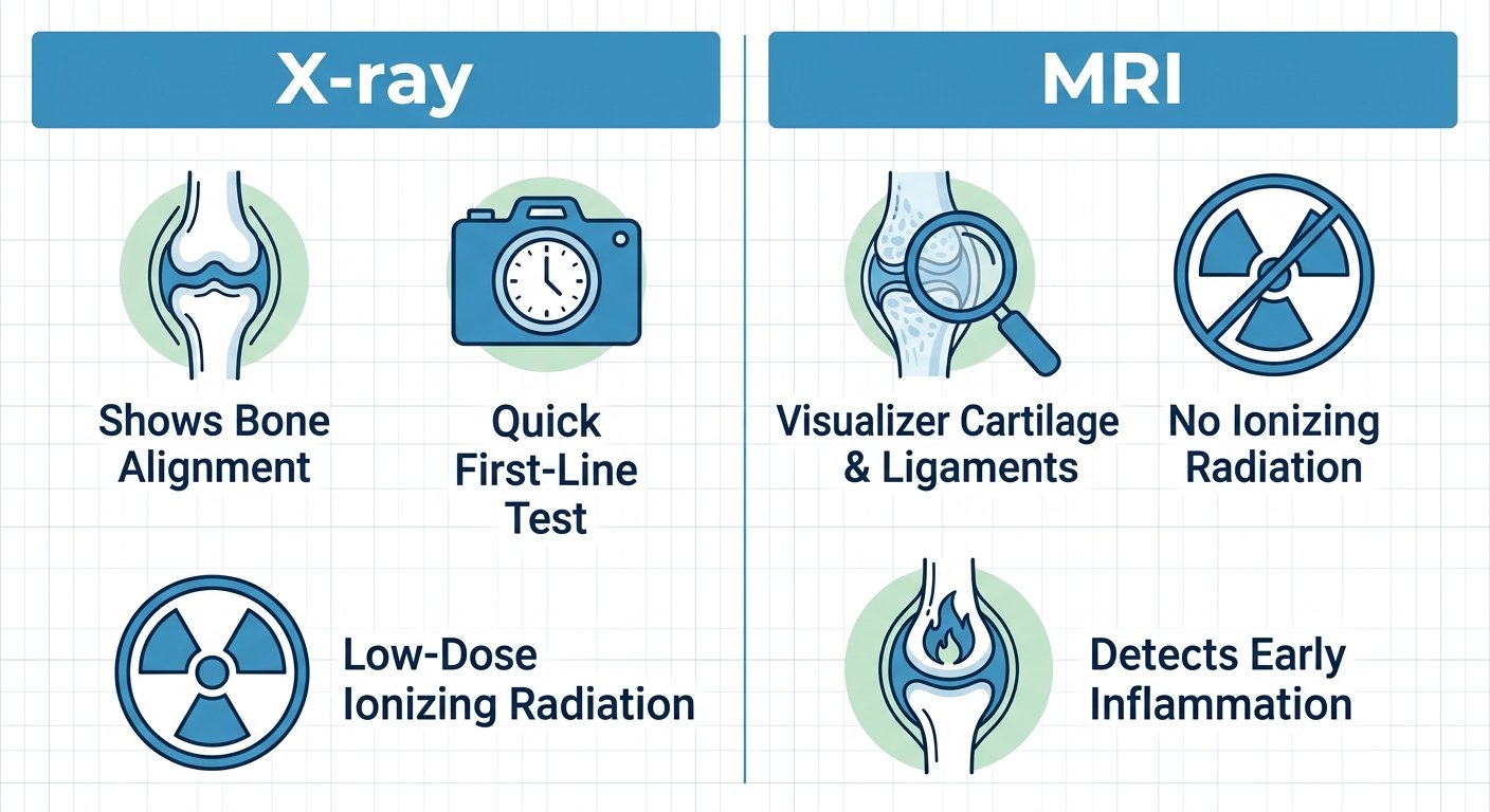
Whether to order an x-ray, MRI or both will be determined by the referring physician based on the patient’s history, physical exam findings, and what structures need to be evaluated. Frequently they may be used together to give the physician more information for diagnosis and treatment.
Beyond X-ray and MRI, other imaging tools such as ultrasound also contribute to arthritis evaluation by detecting joint inflammation and guiding therapeutic injections. Your health and clarity are our priority, and we work closely with referring providers to coordinate the right imaging plan. Please consult your physician to determine which test is appropriate for your situation.
Ultrasound’s Contribution to Diagnosing Inflammatory Arthritis
When considering imaging tests for arthritis diagnosis, ultrasound provides a dynamic and non-invasive method for detecting early signs of inflammatory arthritis, such as synovitis and joint effusion. Unlike X-ray, which primarily visualizes bone, ultrasound can reveal soft tissue changes that often precede permanent joint damage.
One thing ultrasounds can actually measure directly is the state of the membrane layer lining the capsule of the joints, the synovium, which is swollen in synovitis, a component of rheumatoid arthritis as well as other joint conditions. Ultrasound uses high-frequency sound waves to make real-time pictures, demonstrating any increase in the layering thickness, extra fluid collected around the joint (or effusion), plus any signs of early bone erosions that may not yet be visible on radiographs. With Doppler ultrasound, blood flow in the synovium can be evaluated and may indicate disease activity. This functional information helps clinicians gauge disease activity and guide treatment decisions without exposing patients to ionizing radiation.
The dynamic nature of ultrasound further sets it apart from static imaging modalities. The sonographer can examine joints in motion, asking the patient to flex or extend while scanning, which reveals functional abnormalities and subtle synovial recesses that may be missed on still images. Comparing the symptomatic side to the asymptomatic contralateral side adds diagnostic confidence. This hands-on approach also allows for targeted evaluation of tendons, ligaments, and other periarticular structures, providing a comprehensive view of the joint environment.
Because it does not utilize ionizing radiation, ultrasounds can be performed repeatedly in order to track a patient’s disease progress, see how treatment is working, or inject fluid directly into swollen joints. Because of its safety profile, the use of ultrasound as a means to monitor long-term diseases like arthritis is an excellent solution because patients need to have many different images taken over time.
For patients with suspected inflammatory arthritis, ultrasound offers a safe, repeatable, and highly detailed imaging option that supports early diagnosis and ongoing care. At Arizona Advanced Imaging, we provide dedicated musculoskeletal ultrasound using high-frequency transducers, and our board-certified radiologists deliver reports with a 24–48 hour report turnaround, giving referring providers timely, actionable information. The ability to detect active inflammation also helps clinicians differentiate inflammatory arthritis from osteoarthritis, guiding the diagnostic process toward the most appropriate treatment pathway.
Distinguishing Osteoarthritis from Inflammatory Arthritis Through Imaging
Differentiation between osteoarthritis and inflammatory arthritis is critical for deciding treatment, and advanced imaging provides objective evidence when physical examination and laboratory tests leave uncertainty. Osteoarthritis is a chronic condition characterized by the wearing down of cartilage over time. In inflammatory arthritis like rheumatoid arthritis or psoriasis arthritis, the synovial membrane (lining the joints) gets inflamed due to the body’s immune system attacking itself.
Radiographs remain the first-line imaging tests for evaluation of patients with arthritis because they’re readily available, quick to perform, and reveal the bony changes needed to make the diagnosis. Osteoarthritis on X-ray usually shows asymmetrical joint space narrowing, where there was thinning of the cartilage; osteophytes, which represent bony outgrowths along the periphery of the joints; and subchondral sclerosis, a dense area of bone located just underneath damaged cartilage. Inflammatory arthritides show different radiographic findings, such as periarticular osteopenia (bone loss around the joint), marginal erosions due to the erosion of bone from synovial tissue in areas surrounding the joint, and deformity of the affected joint when the disease has been present for many years.
MRI shows important soft-tissue information which the X-ray does not provide. It can reveal the extent of cartilage loss seen in osteoarthrosis; detect a meniscal tear at the knee; show bone-marrow edema in patients suffering from osteoarthrosis, which indicates that mechanical over-load or micro-fracture has taken place in some places within bones. MRI shows synovitis when there exists a thickened, enhancing synovial membrane, shows tenosynovitis affecting the membranes surrounding tendons, and detects bone erosions before they become visible on radiographs in inflammatory arthritis.
Ultrasound delivers real-time imaging without using radiation and is highly sensitive for detecting synovitis as well as the power Doppler signal that indicates active hyperemia in inflamed synovium. For patients with inflammatory arthritis, the degree of Doppler activity may be related to their level of disease activity and help determine treatment options.
It can be used as an add-on if the imaging has complicated anatomy that needs bone detail, like looking at erosions in hands and feet for instance, particularly when other imaging cannot be done due to some reasons such as MRIs being contraindicated. Radiation exposure restricts its usage.
There isn’t always one way to solve a problem by using a single method. Rheumatologists can put their patients’ medical examinations together with radiography test results from different imaging methods to formulate a well-reasoned hypothesis on what may be wrong, and explain how several different tests could be needed when making a diagnosis.
Preparing for Your Arthritis Imaging Appointment and Understanding Your Report
The better you prepare before your arthritis exam, the easier your visit will be, and the clearer any imaging results will be for your doctor to review.
Our staff will coordinate the referral from your referring physician and arrange for a pre-authorization if necessary when you schedule your exam—often with same-day or next-day availability. When making your appointment, bring your insurance card, driver’s license or other valid identification, a copy of the referral order issued by your doctor, a list of current medications, and any prior imaging studies that may be helpful in preparing to complete your examination. We accept almost all major commercial, government and workers’ comp insurance plans and thus our billers work hard to keep costs down as much as possible.
Be at the hospital for the test several minutes prior to start time to complete paperwork. You will be asked to change into a gown. Make sure that jewelry, watches, eyeglasses, hairpins, and any other metal objects are removed, because metal can interfere with the images and affect diagnostic quality. A technologist will explain how we keep you safe from possible harm and will show you what happens during your exam. The technologist also provides instructions for all parts of the exam and answers your questions as well.
Once done scanning, our board-certified radiologists go through your pictures and prepare a detailed report, which usually takes about 24 to 48 hours to process. The report has four major parts.
- Clinical History: The reason for the exam, as provided by your referring physician.
- Technique: The type of imaging performed, such as MRI, CT, X-ray, or ultrasound, and the specific protocols used.
- Findings: A thorough description of what the radiologist observed in the images, noted in precise medical language.
- Impression: A concise summary of the most important observations and possible diagnoses.
In general, the impression section is typically the most actionable part of the report for your referring doctor. While this section highlights the key points that matter for your arthritis management, only your physician can interpret those findings in the context of your complete medical history and physical examination. It’s recommended that you review the entire report together with your referring provider so you understand what the images reveal and what the next steps should be.
Now that we’ve explained our report in detail, you should be ready to confidently chat with your doctor about which imaging tests are best suited for your ongoing care.
Moving Forward: Getting the Right Imaging for Your Arthritis
If you are seeking an arthritis diagnosis, the accuracy depends on the expertise of the radiologist interpreting your images. At Arizona Advanced Imaging, our board-certified, fellowship-trained radiologists specialize in musculoskeletal and neuro imaging, helping to ensure subtle joint changes are not overlooked. We schedule your appointment as convenient for you – either same-day or next day if needed. Once complete, studies are reviewed and reported back usually between 24 and 48 hours to ensure you don’t wait too long. At our imaging centers we accept a wide range of insurance plans—including Medicare, Blue Cross Blue Shield, Cigna, and Aetna—which helps simplify billing and reduce financial stress. After receiving your results, we encourage you to share them promptly with your referring physician to finalize your diagnosis and discuss the best treatment options. With a reliable imaging report in hand, you are ready to move forward with your physician toward a definitive arthritis diagnosis and appropriate treatment plan.
Frequently Asked Questions
Why might a doctor recommend more than one type of imaging test for joint pain?
A doctor may recommend more than one imaging test because each modality highlights different findings. X-rays capture bone alignment and joint space, while MRI reveals cartilage, tendons, and inflammation. Combining them provides a comprehensive view when symptoms suggest both bone and soft-tissue involvement, leading to more precise diagnosis and treatment.
What are the most common imaging tests used to diagnose arthritis?
The most common imaging tests used to diagnose arthritis are X-ray, MRI, CT, and ultrasound. X-ray is typically the first-line tool for bone structure and joint space. MRI excels at soft-tissue detail like cartilage and synovium. CT offers high-resolution bone images, and ultrasound detects synovitis and fluid without radiation.
When should an MRI be considered over an X-ray for arthritis evaluation?
An MRI should be considered over an X-ray when soft-tissue injury or inflammatory arthritis is suspected, or when X-ray results do not explain the patient’s symptoms. MRI visualizes cartilage, ligaments, and bone marrow inflammation that X-ray misses, aiding early detection of conditions like rheumatoid arthritis or meniscal tears.
Can an ultrasound help diagnose inflammatory types of arthritis?
Yes, ultrasound can help diagnose inflammatory types of arthritis. It detects early signs such as synovial thickening, joint effusion, and bone erosions that may not appear on X-ray. Doppler ultrasound can also assess blood flow in the synovium, indicating active inflammation and guiding treatment decisions without radiation exposure.
How do imaging results help differentiate between osteoarthritis and inflammatory arthritis?
Imaging results differentiate osteoarthritis from inflammatory arthritis through distinct findings. Osteoarthritis on X-ray shows asymmetrical joint space narrowing and osteophytes, while inflammatory arthritis presents periarticular osteopenia and marginal erosions. MRI and ultrasound further detect synovitis and soft-tissue inflammation characteristic of inflammatory conditions.
Your Next Steps Toward a Definitive Arthritis Diagnosis
To achieve a definitive diagnosis, many patients turn to imaging tests for arthritis diagnosis, such as MRI, ultrasound, and X-ray. These studies can help your physician confirm the presence and extent of joint involvement. If you suspect arthritis, the first step is to consult your healthcare professional and obtain a referral for the appropriate imaging study. Once you have a referral, you can schedule your appointment at Arizona Advanced Imaging, where we offer advanced open and closed MRI systems, high-frequency ultrasound, and digital X-ray technology tailored for musculoskeletal evaluation. Results may vary based on individual patient conditions, and a healthcare professional should be consulted for diagnosis and treatment advice. Our caring team is ready to assist you and ensure a seamless experience. Call (844) 641-2111 to schedule your imaging study and move toward clearer answers.
Schedule Arthritis Imaging Today
Now that you understand how imaging tests like MRI, X-ray, ultrasound, and CT can distinguish osteoarthritis from inflammatory arthritis, the next step is putting that knowledge into action. Arizona Advanced Imaging offers all of these modalities, interpreted by board-certified, fellowship-trained musculoskeletal radiologists. With same-day and next-day appointments available, you can begin the diagnostic process quickly. Reports are typically delivered within 24 to 48 hours, so you and your physician can move forward confidently. Their team also handles insurance pre-authorization to simplify your experience. Visit our website to learn more or call (844) 641-2111 to schedule your arthritis imaging appointment.
Schedule Arthritis Imaging Today