

Table of Contents
- Understanding Imaging Tests for Prostate Cancer
- The Role of Imaging in Prostate Cancer Diagnosis
- What an MRI Shows for Prostate Cancer
- CT Scans and Other Imaging Modalities for Prostate Evaluation
- Preparing for Your Prostate Imaging Exam
- Interpreting Your Imaging Results and Next Steps
- Your Path to Clear Prostate Health with Arizona Advanced Imaging
Understanding Imaging Tests for Prostate Cancer
Imaging tests for prostate cancer encompass MRI, CT, ultrasound, and nuclear medicine scans that detect tumors, determine cancer stage, and monitor treatment response. These noninvasive exams show the tumor’s location, size, and whether it has spread beyond the prostate, forming the basis for personalized treatment planning. We coordinate with referring physicians to integrate images into individualized care plans.
Accurate imaging guides biopsies to suspicious areas and helps physicians select the most effective intervention, whether active surveillance, surgery, or radiation. This detailed information allows oncologists to tailor treatments with greater confidence, directly impacting patient outcomes. Timely, high-quality imaging reduces repeat exams and helps patients begin appropriate care sooner. At our imaging centers, we use advanced technology to deliver precise results that support both clinical decision-making and patient peace of mind.
Key Takeaways
- Imaging has become indispensable in the evaluation of patients with prostate cancer, particularly in elucidating information that is not obtainable by physical examination.
- Prostate MRI provides high-resolution anatomical detail that helps physicians detect suspicious lesions and assess the likelihood of clinically significant prostate cancer.
- Comprehensive prostate cancer evaluation frequently combines different imaging methods, each answering a distinct clinical question.
- After your prostate MRI exam is complete, a board-certified radiologist carefully analyzes each image and produces a detailed report that communicates the findings to your referring physician.
- Understanding your imaging tests for prostate cancer empowers you to take informed next steps.
The Role of Imaging in Prostate Cancer Diagnosis
Imaging has become indispensable in the evaluation of patients with prostate cancer, particularly in elucidating information that is not obtainable by physical examination. There have been numerous advances in prostate imaging, and it is the particular role (i.e. in the detection, staging and biopsy guidance) that enhances the clinical utility of an imaging modality.
The role of imaging in the workup of the patient with elevated PSA or abnormal digital rectal examination (DRE) is critical. When a patient presents with an elevated prostate-specific antigen (PSA) level or an abnormal digital rectal examination, imaging helps identify suspicious areas within the prostate that warrant closer investigation. In the absence of imaging, the clinician may not identify prostate cancer that is hidden by normal structures.
Beyond detection, imaging is crucial in staging prostate cancer. For instance, imaging helps determine the extent to which prostate cancer has spread to surrounding tissues and whether it has reached lymph nodes or other organs. Imaging results help in determining the overall treatment plan for the patient.
Imaging also transforms how prostate biopsies are performed. Rather than relying solely on systematic, random sampling of the prostate, physicians can use image-guided techniques to direct the biopsy needle to the most suspicious locations. This targeted, image-guided approach improves the precision of tissue sampling and increases the likelihood of capturing clinically significant cancer on the first attempt.
Taken together, imaging results inform the collaborative decisions that urologists and oncologists make with their patients. The findings help determine whether a plan of active surveillance, surgery, or other management strategies is appropriate, though imaging alone never replaces a thorough clinical evaluation or the definitive information provided by a tissue biopsy. It is a powerful tool, but always a part of a larger diagnostic process.
One of the most influential imaging tools in this pathway is magnetic resonance imaging (MRI), which offers detailed soft-tissue visualization of the prostate and surrounding structures. As your trusted radiology partner, we deliver the high-quality MRI and supporting imaging services that healthcare teams rely on when navigating these complex diagnostic decisions.
What an MRI Shows for Prostate Cancer
Prostate MRI provides high-resolution anatomical detail that helps physicians detect suspicious lesions and assess the likelihood of clinically significant prostate cancer. This advanced imaging tool plays a central role in evaluating the prostate gland, local staging of known cancer, monitoring treatment effectiveness, and checking for recurrence.
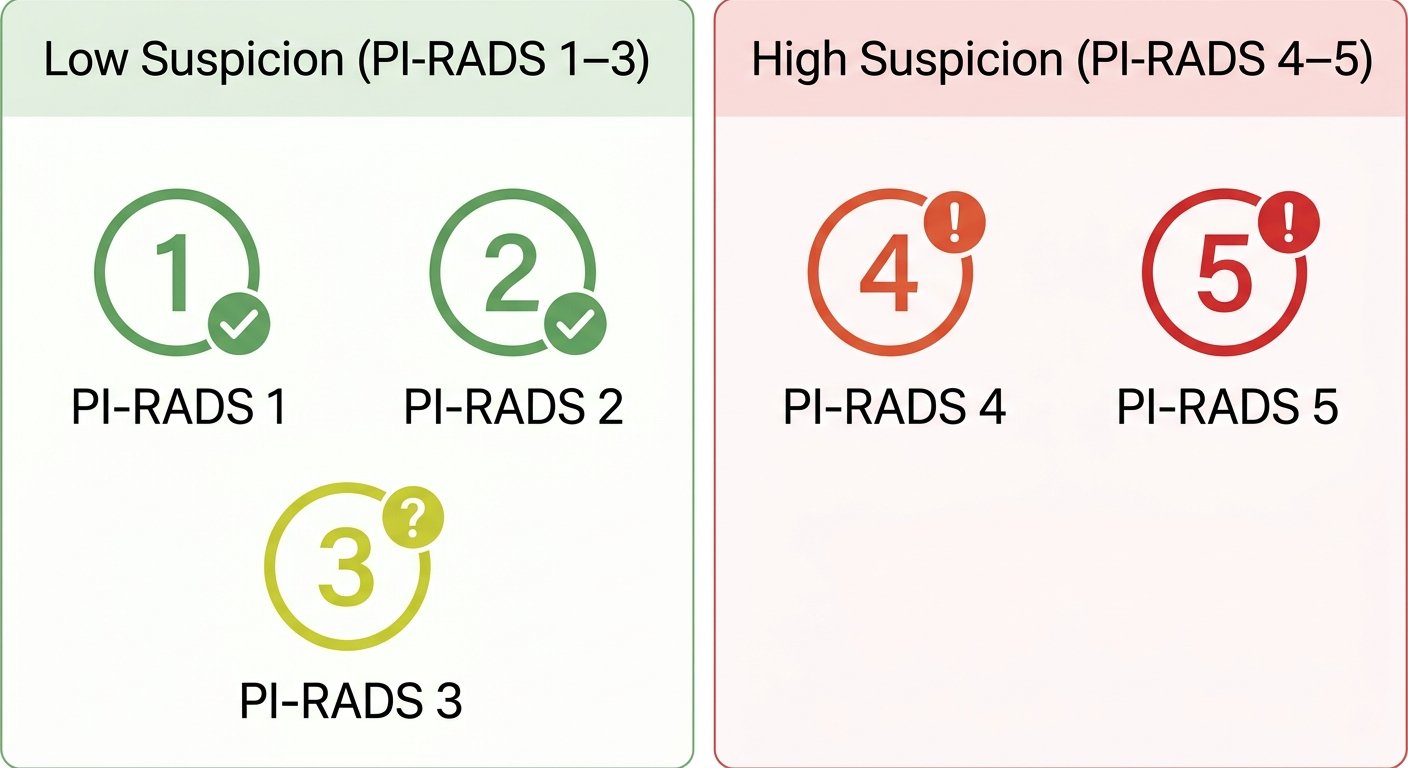
Central to prostate MRI interpretation is the Prostate Imaging Reporting and Data System, or PI-RADS, a standardized 5-point scoring method that communicates the probability that a finding represents clinically significant cancer. According to the Radiological Society of North America’s patient education resource, RadiologyInfo.org, PI-RADS categories range from very low to very high likelihood of clinically significant prostate cancer. A PI-RADS 1 score indicates a very low likelihood, while PI-RADS 2 reflects a low likelihood. PI-RADS 3 represents an intermediate or undetermined probability. PI-RADS 4 indicates a high likelihood, and PI-RADS 5 signals a very high likelihood of clinically significant cancer. These scores guide referring physicians but do not provide a definitive diagnosis — they are interpreted alongside other clinical information such as PSA levels, patient history, and biopsy results.
A standard prostate MRI report typically includes several key sections: the exam type, clinical history, technical details about the imaging process, the radiologist’s findings, and a final impression. The Impression section is particularly critical because it synthesizes the most significant observations for the referring physician, integrating MRI results with the patient’s broader clinical picture. RadiologyInfo.org emphasizes that this section distills what matters most for next steps, including any recommended follow-up imaging or biopsy correlation. Understanding the report structure can help patients have conversations with their healthcare providers about what an MRI shows for prostate cancer and how those findings shape the path forward.
In addition to MRI, other imaging modalities such as CT scans may be used for a fuller picture when evaluating prostate cancer, particularly in assessing whether disease has spread beyond the prostate. Your health and clarity are our priority, and we are committed to producing precise, timely reports that support both clinical care and informed patient discussions.
CT Scans and Other Imaging Modalities for Prostate Evaluation
Comprehensive prostate cancer evaluation frequently combines different imaging methods, each answering a distinct clinical question. CT scans serve a critical role in detecting lymph node involvement and distant metastases, complementing the detailed soft-tissue visualization that MRI provides for the prostate itself. When a prostate cancer diagnosis is suspected or confirmed, physicians often rely on CT for whole-body staging to decide whether additional interventions are necessary.
CT imaging uses a rotating X-ray source to capture cross-sectional images of the chest, abdomen, and pelvis. This wide anatomical coverage is ideal for identifying enlarged lymph nodes, bone metastases, and organ involvement beyond the prostate gland. At Arizona Advanced Imaging, we use low-radiation CT scanners designed to minimize patient exposure while still producing high-resolution diagnostic images. Most CT exams are completed in under 15 minutes, and our board-certified radiologists typically provide a finalized report within 24 to 48 hours. While MRI remains the preferred tool for local prostate staging due to its superior soft-tissue contrast, CT excels at rapidly screening the entire body for distant spread—a key factor in treatment planning.
Other imaging modalities each offer unique insights during prostate evaluation:
- Multiparametric ultrasound uses sound waves to assess prostate size, vascularity, and tissue stiffness without ionizing radiation. It is often employed for biopsy guidance but has limited sensitivity for small metastases.
- PET/CT with PSMA tracers merges functional and anatomical data, enabling highly sensitive detection of prostate cancer cells. This hybrid scan is increasingly used to locate recurrent disease and guide salvage therapies.
- Bone scans detect areas of increased bone turnover, flagging skeletal metastases that may not yet be visible on CT. They remain a common next step when PSA levels rise significantly.
Each tool serves a specific purpose, and the choice depends on the clinical question, disease stage, and patient history.
Clear imaging begins with proper exam preparation. For CT scans, this may involve fasting, drinking oral contrast to highlight the bowel, or temporarily pausing certain medications—all steps our team coordinates in advance to ensure a smooth experience and diagnostic accuracy. The next section explains exactly how to prepare for your imaging appointment so that every study yields reliable, actionable results.
Preparing for Your Prostate Imaging Exam
After understanding the different imaging options available for prostate evaluation, the next practical step is knowing how to prepare for your specific exam. While every study is tailored by your referring physician, a few universal steps help ensure your appointment moves smoothly and your images are of the highest quality.
What to bring
- Insurance card and a photo ID
- The physician referral form, if your plan requires one
- A list of all current medications, allergies, and any prior reactions to contrast agents
- Any previous prostate imaging reports or CDs you have been given
- Comfortable, metal-free clothing (avoid zippers, snaps, and underwire)
- A form of payment for any co-pay or deductible, if applicable
If it is determined that intravenous contrast is needed, you may be asked to avoid food or drink for a few hours beforehand. Our scheduling staff will also verify any implants, pacemakers, or metal fragments that could affect an MRI.
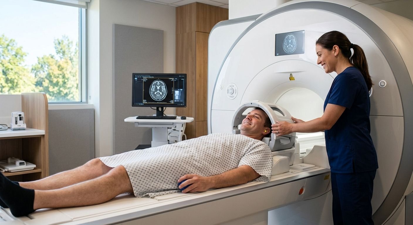
The studies are completely painless and non-invasive. During an MRI, you will hear loud knocking or tapping sounds. These are normal and just indicate that the magnet is working. We offer earplugs to make the experience more comfortable. You may experience a brief warm flushed sensation if a contrast study is done. This is normal and should not cause you any concern. That sensation is expected and harmless and will pass quickly.
Let our staff know when making your appointment if you have claustrophobia or anxiety, and we can make special arrangements for you on a spacious open MRI scanner. You will remain in contact with the MRI technologist via an intercom throughout the exam, and you are able to pause the exam at any time by simply raising your hand. Prostate MRIs generally take 30-60 minutes, and the length of your exam is based on the type of MRI images your doctor has requested.
Once your exam is complete, the images will be interpreted by a board-certified radiologist, and the findings will help guide your next clinical steps. Your physician will receive the report quickly, and our staff will make sure you have a seamless experience from check-in to follow-up.
Interpreting Your Imaging Results and Next Steps
After your prostate MRI exam is complete, a board-certified radiologist carefully analyzes each image and produces a detailed report that communicates the findings to your referring physician. Understanding the key parts of this report can help you have a more informed conversation about your health.
A patient education resource from the American College of Radiology and the Radiological Society of North America notes that a standard prostate MRI report includes six essential sections. The exam type identifies the scan performed. The clinical history summarizes the reason your doctor ordered the imaging. A comparison section references any previous studies for context. The technical details describe how the scan was acquired. The findings provide the radiologist’s observations, including the location and characteristics of any abnormal areas. The final impression synthesizes all of this information into a concise summary that your referring physician uses to determine next steps.
Central to the findings is the Prostate Imaging Reporting and Data System (PI-RADS), a standardized scale that rates the likelihood of clinically significant prostate cancer:
- PI-RADS 1: Very low likelihood
- PI-RADS 2: Low likelihood
- PI-RADS 3: Intermediate (undetermined) likelihood
- PI-RADS 4: High likelihood
- PI-RADS 5: Very high likelihood
The impression section is written specifically for your doctor, who interprets the PI-RADS score alongside your personal medical history and any other test results. It is not a standalone diagnosis; rather, it guides the clinical decisions that you and your physician will make together, such as whether to pursue a biopsy, active surveillance, or additional imaging.
We recommend reviewing the full report with your referring physician, who can explain the implications in the context of your overall health. At Arizona Advanced Imaging, our board-certified radiologists, fellowship-trained in musculoskeletal and neuroradiology, interpret every prostate imaging study. We deliver these reports with a typical turnaround of 24 to 48 hours, so you and your doctor can move forward without delay. With a clear understanding of your imaging results, you can confidently enter the next phase of care, and our team is ready to support you with expert interpretation and rapid communication. To discuss your report or schedule follow-up imaging, call us at (844) 641-2111 or contact our Tempe office in Arizona.
Your Path to Clear Prostate Health with Arizona Advanced Imaging
Understanding your imaging tests for prostate cancer empowers you to take informed next steps. A service of the RSNA and ACR states that the Impression section of your prostate MRI report synthesizes all findings and assigns a PI-RADS score, a 1-to-5 scale that indicates the likelihood of clinically significant cancer and guides your physician’s decision between biopsy and active surveillance.
At our Phoenix-area locations, our board-certified, fellowship-trained radiologists and accredited imaging centers ensure every prostate MRI is read with the expertise and accuracy patients and referring doctors need to move forward with confidence. We provide prompt, patient-focused support — from pre-authorization assistance to 24-to-48-hour report turnaround.
Take the next step: schedule your prostate MRI by calling (844) 641-2111 or by visiting our Phoenix office at 1775 W University Dr, Suite 127, Tempe, AZ 85281. Results may vary based on individual patient conditions. Please consult with a healthcare professional for diagnosis and treatment advice.
Schedule Your Prostate MRI
You have explored how imaging tests for prostate cancer, including MRI with PI-RADS scoring, provide essential information for diagnosis and treatment planning. The Impression section of your report synthesizes findings to guide your physician’s next steps. In Tempe, our board-certified radiologists deliver precise, timely interpretations you can trust. Our commitment to rapid 24-48 hour reporting ensures you receive results without unnecessary delay. Now that you understand the value of expert imaging, it’s time to take action. Schedule your prostate MRI appointment today and move forward with confidence.