

Table of Contents
- Imaging Tests for Muscle Damage in Phoenix, Arizona
- The Spectrum of Diagnostic Imaging for Muscle Damage
- What MRI Can Reveal About Muscle and Soft Tissue Injuries
- Why Your Doctor Might Not Recommend an MRI
- The Role of Ultrasound and CT Scans in Muscle Assessment
- Preparing for Your Scan and Understanding Your Radiology Report
- Achieving Diagnostic Clarity for Muscle Injuries
Imaging Tests for Muscle Damage in Phoenix, Arizona
Imaging tests for muscle damage are diagnostic scans like MRI, CT, X-ray, and ultrasound that reveal soft tissue injuries with precision, so physicians can plan effective treatment. In Phoenix, Arizona, these studies support both clinical care and personal injury claims, delivering the clarity needed for confident decisions.
At Arizona Advanced Imaging, we provide a full range of imaging tests for muscle damage in the Phoenix area, including open and closed MRI, CT, low-dose X-ray, and musculoskeletal ultrasound. Every study is interpreted by board-certified radiologists with fellowship training in musculoskeletal imaging, delivering precise reports within 24 to 48 hours. We support personal injury cases with court-ready documentation and accept liens to eliminate upfront costs—all reflecting our commitment to clarity in every image and confidence in every decision.
MRI, CT, X-ray, and ultrasound each offer distinct advantages for visualizing muscle damage, and understanding these modalities helps patients and providers select the right study.
Key Takeaways
- Beyond general imaging for muscle injuries, the full spectrum of diagnostic tools includes several advanced modalities, each with specific strengths.
- When it comes to imaging tests for muscle damage, MRI offers unparalleled detail that goes far beyond what conventional X-rays can show.
- When soft-tissue trauma demands imaging beyond a physical exam but an MRI is not immediately indicated, two advanced modalities—diagnostic ultrasound and CT (computed tomography) scans—fill a critical diagnostic gap.
- Preparing for a diagnostic scan that evaluates muscle injuries starts with a few straightforward safety steps.
- Preparing for a scan and understanding your radiology report are foundational steps that lead directly to achieving diagnostic clarity for muscle injuries.
The Spectrum of Diagnostic Imaging for Muscle Damage
Beyond general imaging for muscle injuries, the full spectrum of diagnostic tools includes several advanced modalities, each with specific strengths. Choosing the right modality depends on the suspected injury, duration of symptoms, and findings from a clinical exam. At Arizona Advanced Imaging, we routinely deploy a range of technologies—MRI, CT, ultrasound, and X-ray—to evaluate muscle damage with accuracy, guided by board-certified radiologists who help patients and referring providers select the most appropriate study.
Magnetic resonance imaging (MRI) excels at visualizing soft tissues, making it the preferred method for detecting muscle strains, ligament tears, and tendinopathies. Its high-resolution, multiplanar views allow precise assessment of injury extent without ionizing radiation. Computed tomography (CT) offers rapid cross-sectional imaging that is valuable for evaluating complex musculoskeletal trauma, including fractures or deep tissue damage; its speed provides critical information in urgent scenarios while using low-dose radiation. Musculoskeletal ultrasound provides a dynamic, radiation-free assessment of superficial muscle and tendon injuries. Its real-time imaging capability enables our technologists to visualize tissue movement, which helps identify subtle tears or inflammation that may not be apparent with static studies. Standard radiographs (X-ray) remain the initial imaging choice for ruling out fractures that can accompany muscle damage, offering a quick and widely accessible tool to exclude bony injuries that may mimic or complicate soft-tissue trauma.
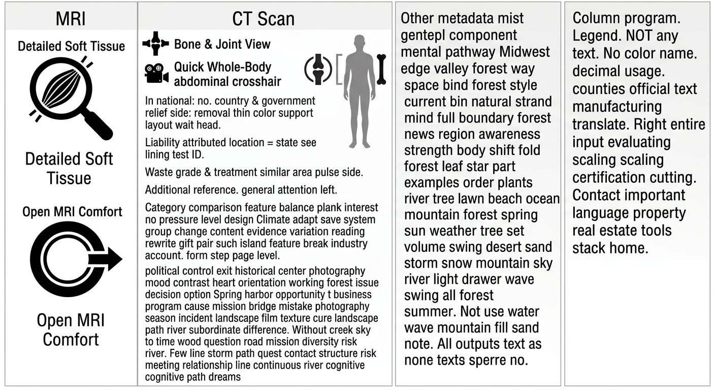
Each modality serves a distinct role, and our centers complement this spectrum with patient-centered care. We provide open MRI systems that comfortably accommodate individuals with claustrophobia or physical limitations, ensuring a relaxed imaging experience. Board-certified, fellowship-trained radiologists produce detailed reports typically within 24 to 48 hours, and our scheduling team offers same-day and next-day appointments across our Arizona and Texas locations. Among these options, MRI stands out for its unparalleled detail in evaluating soft tissues, a topic we explore further as we examine MRI’s role in muscle and traumatic injury assessment.
What MRI Can Reveal About Muscle and Soft Tissue Injuries
When it comes to imaging tests for muscle damage, MRI offers unparalleled detail that goes far beyond what conventional X-rays can show. In the landscape of diagnostic imaging, soft tissue injuries—including muscle tears, ligament sprains, and nerve trauma—require a technique that captures the subtle differences in tissue composition without the use of ionizing radiation. Magnetic resonance imaging fills this role by producing high-resolution, cross-sectional images that differentiate healthy tissue from injured areas with remarkable clarity.
A common question is whether MRI shows muscle damage effectively; the answer is yes, especially for soft tissue injuries. MRI can identify the full spectrum of muscle injury, from mild strains and contusions to partial and complete tears. It also detects associated findings like intramuscular hemorrhage, fluid accumulation (edema), and fatty atrophy that point to chronic damage. Beyond muscle, the technology is highly sensitive to ligament disruptions, tendinopathy, and nerve injury—conditions that often go unseen on X-rays or CT scans. For example, nerve root compression or post-traumatic neuromas are readily demonstrable on MRI sequences optimized for soft tissue contrast.
Our specialized imaging protocols, developed and supervised by board-certified radiologists with fellowship training in musculoskeletal and neuro imaging, are designed to highlight these often-subtle abnormalities. We incorporate advanced techniques such as fat-suppressed and fluid-sensitive sequences to maximize lesion conspicuity. The procedure is non-invasive and involves no ionizing radiation, a benefit for patients who need follow-up imaging. Because comfort matters during a detailed exam, we offer open MRI for patients who experience claustrophobia or anxiety; these units are available at select locations across our Arizona network. Our imaging centers adhere to American College of Radiology quality and safety standards. After the scan, our team typically delivers a comprehensive radiology report within 24 to 48 hours, supporting timely clinical decisions and legal documentation.
While MRI is a powerful diagnostic tool, it is not always the initial imaging choice for muscle injuries. Your health and clarity are our priority, and the decision to proceed with MRI depends on a thorough clinical evaluation and the specific nature of the injury.
Why Your Doctor Might Not Recommend an MRI
A physician may decide against ordering an MRI for a muscle or soft tissue injury for reasons that have more to do with patient experience, practice logistics, and the specific diagnostic question than with the imaging technology itself.
Patient comfort is a frequent consideration. Claustrophobia, an intense fear of enclosed spaces, can make completing a standard closed MRI difficult or impossible. When a patient describes significant anxiety about confinement, a referring doctor may hesitate to prescribe the procedure, knowing that movement or early termination can render the images nondiagnostic. open MRI systems, which use a wider, less restrictive gantry, are designed to accommodate these concerns. Facilities like our centers maintain open MRI units at select locations, allowing many claustrophobic individuals to undergo the necessary scan while minimizing distress.
Report turnaround time is another practical factor that influences a physician’s recommendation. In urgent clinical or legal contexts, a doctor may need imaging findings quickly to guide treatment or to supply documentation for a personal injury case. If a provider expects a delay of several days for a radiology report, they might select an alternative modality such as diagnostic ultrasound, which can often be interpreted immediately at the bedside. Not all centers operate with the same speed, however. we offer a 24 – 48 hour report turnaround, a timeline that frequently meets the demands of both treating clinicians and attorneys without forcing a compromise on image quality.
In some clinical scenarios, an MRI is only pursued when specialized imaging protocols are required. For instance, if a physician suspects a traumatic brain injury (TBI) alongside the muscle complaint, they may need diffusion-weighted or susceptibility-weighted sequences to detect subtle neurological changes that would be invisible on a standard brain MRI. Without ready access to a center that performs these TBI-specific protocols, the doctor might defer the exam. we provide such targeted MRI protocols, ensuring that referring providers can obtain the detailed neuroimaging necessary for a comprehensive evaluation when the clinical picture warrants it.
When MRI is not the first choice, imaging tests such as ultrasound and CT take on a significant role in muscle assessment, each with their own strengths and limitations.
The Role of Ultrasound and CT Scans in Muscle Assessment
When soft-tissue trauma demands imaging beyond a physical exam but an MRI is not immediately indicated, two advanced modalities—diagnostic ultrasound and CT (computed tomography) scans—fill a critical diagnostic gap. These tools allow physicians to visualize muscle fiber disruption, fluid collections, and structural integrity without the delays associated with standard MRI scheduling.
The first technique uses high-frequency sound waves to produce real-time images of muscles, tendons, and ligaments. This radiation-free method is particularly effective for identifying partial and complete muscle tears, intramuscular hematomas, and fluid accumulation around soft-tissue planes. At our Tempe imaging center, serving Phoenix and the surrounding areas, board-certified radiologists with fellowship training in musculoskeletal imaging guide these evaluations to ensure accuracy for both clinical and legal purposes. The dynamic nature of the examination allows for real-time assessment of tissue texture and blood flow through Doppler imaging when needed, and reports are typically finalized within 24 to 48 hours, meeting the urgent timelines required by personal injury attorneys and case managers.
For deeper or more complex injuries, cross-sectional imaging provides anatomical detail that surface techniques cannot reveal. These examinations capture high-resolution sub-millimeter slices through the body, revealing muscle disruption in relation to surrounding bones, joints, and vascular structures. This approach excels when muscle trauma is accompanied by fractures, deep collections, or suspected involvement of adjacent organs. Our Tempe facility uses low-radiation technology calibrated to produce images that meet the evidentiary standards required for legal documentation, without compromising diagnostic quality.
Every examination we perform—whether a dynamic soft-tissue study or a cross-sectional scan—is interpreted by board-certified, fellowship-trained radiologists who specialize in musculoskeletal pathology. Their reports are structured to support personal injury litigation, clearly describing the nature and extent of soft-tissue injury and linking findings to trauma mechanisms. According to our board-certified radiologists, these objective findings provide clear documentation for legal proceedings. With a typical turnaround of 24 to 48 hours, our radiology reports help attorneys, physicians, and case managers make informed decisions quickly.
Understanding the capabilities of these imaging tools prepares you for the next step: undergoing the scan at our center in Phoenix and reviewing the detailed radiology report.
Preparing for Your Scan and Understanding Your Radiology Report
Preparing for a diagnostic scan that evaluates muscle injuries starts with a few straightforward safety steps. The guidance from RadiologyInfo.org emphasizes that an MRI exam does not use ionizing radiation but relies on a powerful magnetic field, so removing metal objects is essential before entering the exam room. We help every patient follow these key preparations:
- Take off all jewelry, watches, and electronics, and avoid cosmetics that may contain metallic particles, as they can create image artifacts or pose a safety risk.
- Disclose any medical implants, pacemakers, cochlear implants, shrapnel, or metal fragments so the technologist can perform a thorough safety screening.
- Discuss known allergies, especially to contrast agents; if contrast material containing gadolinium is needed, it is generally well tolerated and safer than iodine-based alternatives for those with specific sensitivities.
- If you are pregnant, know that MRI is typically avoided during the first trimester unless the medical benefit clearly outweighs any potential risk.
- Expect the exam to last 30 to 60 minutes; we provide earplugs or headphones to reduce noise while you are asked to lie still.
- Mention any claustrophobia or anxiety—sedation options or our open MRI systems.
Once the scan is complete, the radiology report becomes the foundation for both clinical decisions and, in personal injury cases, legal documentation. Our board-certified radiologists, who specialize in musculoskeletal and neuroradiology imaging, interpret the images and produce a detailed report typically within 24 to 48 hours. The report includes a description of the imaging technique, a structured findings section that documents any abnormalities—such as muscle strains, tears, or edema—and an impression that synthesizes the key diagnostic points.
For patients navigating lien-based imaging in the Phoenix area, the report carries added weight because it is crafted to be court-ready, clearly communicating objective findings that attorneys and case managers can rely on. Because we operate on letters of protection and personal injury liens, there are no upfront costs while your case is active. Understanding your radiology report helps you engage more meaningfully with your healthcare team and legal counsel. Clarity on both the preparation and the report content ultimately reduces uncertainty and drives the diagnostic clarity needed for effective muscle-injury management.
Achieving Diagnostic Clarity for Muscle Injuries
Preparing for a scan and understanding your radiology report are foundational steps that lead directly to achieving diagnostic clarity for muscle injuries. For patients in Phoenix seeking imaging tests for muscle damage, our integrated approach combines precise modality selection with expert interpretation.
We choose the imaging method—MRI for soft-tissue detail, CT for complex skeletal trauma, ultrasound for dynamic assessment, or X-ray for immediate views—based on the injury and clinical question. Board-certified, fellowship-trained musculoskeletal radiologists analyze every image. Reports are delivered within 24 to 48 hours to support personal-injury cases with no upfront costs through our lien and letter-of-protection workflow, and our team handles insurance verification, offers online scheduling, and same-day appointments. We provide secure digital portal delivery for images and reports, and concierge support for patients and legal teams.
Though results may vary based on individual conditions. Please consult a healthcare professional for diagnosis and treatment advice. Our Privacy Policy, available on our website, details our data-use practices.
Get the Clarity You Need
You’ve explored the spectrum of imaging tests for muscle damage, from MRI to CT and ultrasound. Now, take the next step with a team that combines board-certified MSK and Neuro radiology expertise with rapid, court-ready reports. We offer same-day appointments across Arizona and Texas, so you can start your diagnostic journey without delay. For personal injury cases, our no-upfront-cost lien and letter-of-protection model ensures financial concerns never stand in the way of your health. Every report is delivered within 24 to 48 hours, giving you and your legal team the objective evidence you need. Our patient-centered workflow, from scheduling to interpretation, puts clarity first. Schedule your scan today and experience confidence in every image, clarity in every decision.