

Table of Contents
- A Comprehensive Guide to the CBI-M Traumatic Brain Injury Framework
- Foundations of Traumatic Brain Injury Classification and the CBI-M Model
- Imaging and Biomarker Integration: The Core Pillar of the CBI-M Framework
- Implementing the CBI-M Framework in Clinical and Legal Settings
- Quantitative DTI and SWI: Deepening Diagnostic Precision
- Common Questions About CBI-M and TBI Imaging
- Elevating TBI Care with the CBI-M Framework
A Comprehensive Guide to the CBI-M Traumatic Brain Injury Framework
The CBI-M traumatic brain injury framework is a structured classification system that helps clinicians and imaging teams understand the type and severity of traumatic brain injuries. We rely on such classification to guide our imaging recommendations—from open MRI scans to specialized TBI protocols—so that physicians and legal teams receive reliable, objective data. We explain what the CBI-M framework is, how it organizes brain injury categories, and why that structure leads to more targeted imaging choices.
We provide a basic overview of TBI classification, cover the key elements of the CBI-M model, and describe the application of advanced MR imaging protocols (such as the SpinTech™ MRI TBI Protocol) to identify microhemorrhages and alterations of white matter in the brain. This document is intended for the general audience, e.g. patients and/or their family members evaluating their imaging options, and/or attorneys and law firms evaluating potential personal injury claims.
We acknowledge the importance of clinical and/or subjective assessment and complementary imaging in the evaluation of TBI. Our commitment to clarity in every image and confidence in every decision flows from this classification-first mindset. Therefore, in line with our classification-first strategy, we will cover the basic elements of TBI classification and the place of CBI-M before describing the imaging options.
Foundations of Traumatic Brain Injury Classification and the CBI-M Model
Traditionally, traumatic brain injuries (TBIs) are classified as mild, moderate, or severe, often using the Glasgow Coma Scale (GCS) to measure severity. A patient scores mild TBI when their GCS score is 13 to 15. A patient scores moderate TBI when their GCS score is 9 to 12. A patient scores severe TBI when their GCS score is 8 or less. Duration of loss of consciousness and length of post-traumatic amnesia are added to sharpen the severity picture.
Two patients with the same GCS score may show different symptoms, injury patterns, and recovery rates. The majority of TBIs are classified as mild. Often, conventional Computed Tomography (CT) scans or Magnetic Resonance Imaging (MRIs) do not show abnormalities, and patients are left to manage persistent post-concussive symptoms.
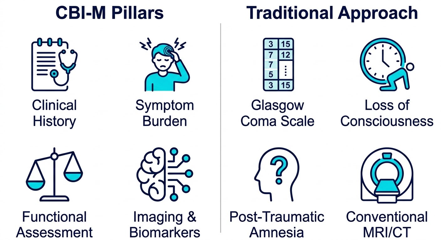
Trying to fill that gap, the CBI-M model uses a multi-pillar assessment instead of a single assessment. The four pillars of the model are: Clinical History, Symptom Burden, Functional Assessment, and Imaging & Biomarkers.
| Assessment Component | CBI-M Framework | Traditional Approach | Clinical Advantage |
|---|---|---|---|
| Clinical History | Detailed accident history, prior concussions, and comorbidities | Loss of consciousness duration | Adds context beyond a single acute variable |
| Symptom Burden | Type, severity, and persistence of symptoms via validated checklists | Post-traumatic amnesia duration | Reflects daily patient experience, not just one parameter |
| Functional Assessment | Cognitive, balance, and oculomotor testing post-injury | Glasgow Coma Scale | Detects subtle functional deficits GCS cannot measure |
| Imaging & Biomarkers | Advanced MRI (quantitative DTI, volumetric analysis, microbleed detection) | Conventional CT or MRI | Reveals microstructural injury invisible to routine scans |
Functional assessment may uncover balance or processing-speed deficits that significantly affect daily life while routine imaging appears unchanged.
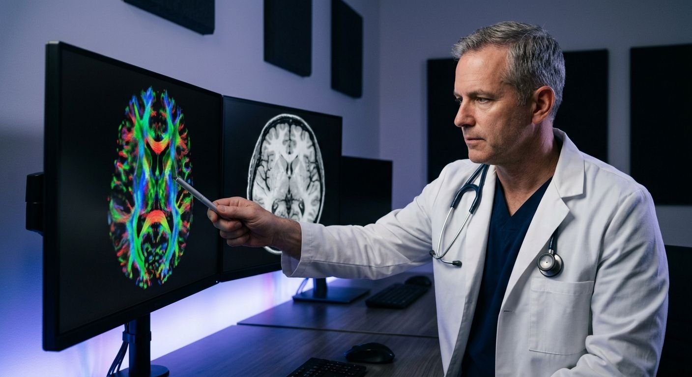
The Imaging & Biomarkers pillar serves as the model’s objective anchor. Our centers utilize the SpinTech MRI TBI protocol, an advanced imaging sequence, which incorporates quantitative diffusion tensor imaging, and is able to detect microstructural changes that may be missed in standard sequences. As your trusted radiology partner, we provide this protocol throughout Arizona and Texas. Reports, reviewed by a fellowship-trained neuroradiologist, are provided within 24-48 hours.
Radiology, therefore, becomes the objective, quantifiable anchor for clinical and symptom burden and functional performance. For patients and their referring clinicians, we encourage imaging finding review with a qualified physician to aid in placement of the imaging findings in the context of symptoms and to assist with care planning.
Imaging and Biomarker Integration: The Core Pillar of the CBI-M Framework
The CBI-M framework is built upon objective, tissue-level evidence. Advanced imaging provides that layer. Unlike assessment tools that depend on patient self-report or bedside observation alone, imaging-derived biomarkers record injury directly from brain tissue.
The Role of Advanced MRI and CT in CBI-M
Standard emergency CT and conventional MRI protocols were not designed to capture the microstructural and microvascular injuries that define subacute and chronic traumatic brain injury. The CBI-M framework addresses this gap by integrating advanced MRI sequences, diffusion tensor imaging, or DTI, which measures the integrity of white-matter tracts by tracking water molecule movement, and susceptibility weighted imaging, which amplifies the magnetic signal of blood products to reveal microhemorrhages — along with CT when indicated.
Our TBI-specific protocol exemplifies how dedicated imaging protocols strengthen the CBI-M evidence layer. The FDA-cleared equipment allows for quantitative DTI and volumetric analysis of the brain, as well as for the automated detection of microbleeds. Conventional MRI may miss up to 50 percent of cerebral microbleeds present in mild TBI cases, while this protocol is backed by more than 100 published research studies supporting its diagnostic sensitivity. Fellowship-trained, board-certified neuroradiologists interpret the studies and create structured reports across our network of centers in Arizona and Texas.
How Quantitative Imaging Enhances Mild TBI Diagnosis
The CBI-M framework alters the diagnostic equation to incorporate quantitative DTI (diffusion tensor imaging) metrics (i.e. fractional anisotropy and mean diffusivity) to describe the directional movement of water molecules along axonal pathways, as well as susceptibility-weighted imaging signal voids, which represent sites of parenchymal hemorrhage. When combined with sequences that show susceptibility-weighted imaging microhemorrhages and tract-based DTI analysis, this framework defines a multidimensional injury profile to classify patients where the Glasgow Coma Scale alone would suggest no significant abnormality.
CBI-M vs. Glasgow Coma Scale: Why Multidimensional Wins
The GCS ranges from 3 to 15 and relies entirely on eye-opening, verbal, and motor responses — behaviors that are easily confounded by intubation, sedation, intoxication, or pre-existing aphasia. In mild TBI, patients frequently score near the top of that range, yielding little clinical differentiation even when imaging shows microstructural injury.
| Feature | CBI-M | Glasgow Coma Scale |
|---|---|---|
| Primary assessment method | Multidimensional: advanced MRI/CT, quantitative biomarkers, and clinical history | Unidimensional: numeric 3–15 score from eye, verbal, and motor responses |
| Detects microstructural injury | Yes — DTI and susceptibility weighted imaging reveal white-matter damage and microhemorrhages | No — the score does not assess brain tissue directly |
| Sensitivity in mild TBI | High — captures injury patterns standard scans miss | Low — frequently normal in mild or concussive injury |
| Prognostic value | Strong — imaging biomarkers inform recovery trajectory and long-term monitoring | Limited — the acute score correlates modestly with long-term outcome |
| Documentation value for PI/legal cases | Strong — objective imaging record supports baseline and serial comparison | Weak — subjective and time-dependent, influenced by sedation or intubation |
Our radiologists provide court-ready reports to quantify tissue-level findings, which provide referring attorneys with objective evidence based on measured data rather than bedside evaluation.
CBI-M anchors the framework in quantitative imaging data, allowing for a baseline against which to measure, assess, and classify the impact of the injury. It guides rehabilitation and support clinical follow-up, as well as documents requirements for litigation.
Implementing the CBI-M Framework in Clinical and Legal Settings
At Arizona Advanced Imaging, we have streamlined our procedures from case referral to the delivery of the report to make the structure’s promise of generating sensitive, court-ready evidence a practical reality for personal injury cases in Arizona and Texas.
Streamlining Workflows for Personal Injury Cases
When a personal injury attorney or physician in Arizona or Texas makes a referral, our process immediately integrates with that of the client. We accept letters of protection and liens, meaning the patient pays nothing out of pocket. Our insurance team obtains pre-authorization for the appointment from commercial, workers’ compensation, and government plans. Our referral process includes the following key steps:
- Referral and Financial Clearance – We confirm the lien or letter of protection and verify insurance details, ensuring no out-of-pocket cost for the patient.
- Appointment Scheduling – Same-day or next-day appointments are booked online or by phone at one of our more than eight imaging centers in Arizona and Texas.
- Pre-Authorization Completion – Staff secure prior authorization, removing administrative delays.
- Comfortable Imaging – The patient undergoes a TBI-specific MRI protocol, including this TBI protocol, on an open or closed MRI system.
- Expert Review – A board-certified, fellowship-trained neuroradiologist interprets the quantitative DTI and SWI data.
- Rapid Report Delivery – The finalized radiology report is ready within 24 to 48 hours and shared securely with the legal team and treating providers.
Using DTI and SWI for Forensic-Quality Reports
Quantitative diffusion tensor imaging and susceptibility-weighted imaging produce numerical and visual data that must be translated into language a jury can understand. The following table summarizes the key DTI and SWI parameters and their legal relevance:
| DTI Metric | What It Measures | Injury Indication | Legal Weight |
|---|---|---|---|
| Fractional Anisotropy (FA) | Directional water diffusion in white matter | Reduced FA suggests axonal injury | Strong objective metric; directly quantifiable |
| Mean Diffusivity (MD) | Average magnitude of water diffusion | Elevated MD may indicate edema or degeneration | Corroborative evidence supporting injury |
| Radial Diffusivity (RD) | Diffusion perpendicular to axonal fibers | Elevated RD suggests demyelination | Supports white matter injury claims; objective marker |
| SWI Microhemorrhage Count | Hemosiderin deposits from shear injury | Presence of microhemorrhages consistent with traumatic shear | Strong corroborative evidence; visually identifiable microbleeds |
When we compare a patient’s fractional anisotropy or radial diffusivity against age-matched normative databases, deviations appear as objective markers of white matter compromise. The report then frames such findings using qualifying language—‘consistent with axonal injury’ or ‘suggests demyelination’—avoiding definitive diagnoses while still providing concrete evidence.
Ensuring Rapid Turnaround and Court-Ready Documentation
Our commitment to a 24–48 hour report turnaround means attorneys receive the completed neuroradiology report within two business days of the scan. Every report is reviewed and signed by a board-certified neuroradiologist with fellowship training in trauma imaging, meeting the evidentiary requirements in Arizona and Texas courts. We provide secure, encrypted images to the legal counsel and treating physicians of the patient. Through the use of structured reports, along with DTI and SWI, settlements and litigation are supported.
The quantitative metrics of fractional anisotropy and radial diffusivity, along with SWI microhemorrhage, help provide the confidence and evidentiary weight required for litigation.
Quantitative DTI and SWI: Deepening Diagnostic Precision
The precision of this framework depends on imaging that goes beyond conventional MRI. At Arizona Advanced Imaging, we rely on our TBI MRI protocol, which integrates quantitative DTI and SWI to provide objective, legally defensible data. Quantitative DTI (diffusion tensor imaging) measures the directional movement of water molecules along white matter tracts, yielding metrics like fractional anisotropy (FA) and mean diffusivity (MD) that can reveal axonal injury invisible on conventional structural scans. SWI (susceptibility-weighted imaging) uses magnetic susceptibility differences to detect traumatic microbleeds and hemosiderin deposits, markers of hemorrhagic axonal injury. Together, DTI and SWI offer complementary information: DTI captures microstructural white-matter disruption while SWI identifies hemorrhagic components, producing a more complete view of traumatic brain injury than either sequence alone. This combined approach also supports medicolegal documentation in personal injury cases by providing objective, spatially specific imaging findings.
The following table illustrates how SWI compares with GRE for microbleed detection.
SWI vs. GRE for Traumatic Microbleed Detection
| Feature | SWI | GRE |
|---|---|---|
| Sensitivity to microbleeds | Higher — detects more traumatic microbleeds | Lower — may miss small microbleeds |
| Visualization of hemosiderin | Superior — hemosiderin appears more conspicuous | Moderate — visible but less pronounced |
| Ability to track chronic injury | Better — captures cumulative microbleed burden | Limited — less sensitive for chronic changes |
SWI’s higher sensitivity and superior hemosiderin visualization make it the preferred sequence for documenting chronic microbleed burden in the CBI-M protocol.
These imaging details naturally raise practical questions about how the framework is applied in individual cases, in clinical and legal contexts.
Common Questions About CBI-M and TBI Imaging
SWI and DTI metrics provide additional detail in brain imaging, but patients often ask how the CBI-M framework translates those numbers into meaningful TBI insights.
What does the CBI-M framework do for TBI imaging?
It provides a means of integrating the quantitative metrics of brain imaging with a patient’s clinical narrative.
What can a dedicated TBI MRI show?
Through the use of a specialized MRI protocol with SpinTech MRI technology, we are able to image some types of white matter injury and brain volume changes that are often undetectable using conventional MRI.
Does the CBI-M framework replace a clinical evaluation?
No. The CBI-M framework is a complementary aid that provides objective data, but does not replace a physician’s clinical assessment.
By systematizing the interpretation of brain imaging using the CBI-M framework, clinicians can improve the precision of TBI diagnosis and management.
Elevating TBI Care with the CBI-M Framework
The concerns reflected by the questions above are common. The CBI-M traumatic brain injury framework offers a detailed, advanced-imaging approach to identify morphological changes associated with TBI. AZ Advanced Imaging has developed a specific MRI TBI protocol based on the CBI-M framework. We offer patients convenient, same-day, or next-day appointments, assist with insurance and pre-authorization, and deliver radiology reports within 24 to 48 hours. If you or a patient could benefit from further assessment and imaging for TBI, contact us to schedule an appointment. Advanced TBI imaging may provide further information on previous neurological injuries and changes. Results may vary; please consult with a healthcare professional for diagnosis and treatment advice.
Schedule Your TBI MRI Exam
Now that you’ve explored the CBI-M traumatic brain injury framework, you understand how advanced MRI provides the objective evidence layer for TBI classification. The framework’s imaging pillar leverages quantitative DTI and SWI to reveal microstructural and microvascular injuries that standard scans miss. At Arizona Advanced Imaging, our board-certified neuroradiologists use a dedicated TBI MRI protocol to deliver court-ready, clinically rigorous reports. We typically return finalized reports within 24 to 48 hours, helping reduce delays in treatment and legal proceedings. For personal injury cases, we accept liens and letters of protection, so there are no upfront costs. Take the next step toward a precise evaluation by scheduling your TBI MRI with our team.