

Table of Contents
Bone Density Scans and MRI: Knowing the Difference
Knowing the difference between a bone density scan and an MRI for cancer can help patients from Arizona and Texas pick the correct imaging exam for their needs. A DEXA scan measures bone mineral density (at the lumbar spine and hips) to diagnose osteoporosis and to assess your fracture risk, while bone scans and MRIs examine bone activity as well as soft tissue, especially for workup of cancer cases.
We’ll explain what each test does, why doctors order it, and how your choice matters. Knowing this will empower you and your doctor to make decisions together about your imaging tests.
Key Takeaways
- bone density scanning uses dual-energy X-ray absorptiometry (DEXA/DXA), which sends two low-dose X-ray beams through the bone at different energies.
- A bone density scan looks at the amount of minerals in bone, whereas an MRI goes deeper into the body’s soft tissue, bone marrow and nerves.
- No, a DEXA scan measures bone mineral density at the spine and hips to diagnose osteoporosis and assess fracture risk, but it can’t be used to detect tumors or metastases.
- When we are looking in detail at a specific area, MRI shines because it can produce clear images of both soft tissue and bone marrow.
- Patients who are researching bone density scan vs MRI for cancer may be unsure if a bone scan and a bone density test are the same exam.
1. What a Bone Density Scan Measures
bone density scanning uses dual-energy X-ray absorptiometry (DEXA/DXA), which sends two low-dose X-ray beams through the bone at different energies. The software then subtracts the attenuation of soft tissues to give an accurate measurement of the true bone mineral density.
Lumbar spine and hips are the main measurement sites. Some exams may also include a Vertebral Fracture Assessment to screen for spine deformities. This scan is the standard first imaging test for suspected low bone density and gives clinicians important information before any other type of modality is considered.
Results are presented as T-scores and Z-scores. A T-score of -2.5 or below indicates osteoporosis, while a T-score between -1.1 and -2.4 indicates low bone mass (osteopenia). The amount of radiation exposure during a DXA scan is approximately one-tenth that of a standard chest X-ray, ensuring safety for the patient while providing accurate diagnostic information.
2. What an MRI Shows for Cancer Care
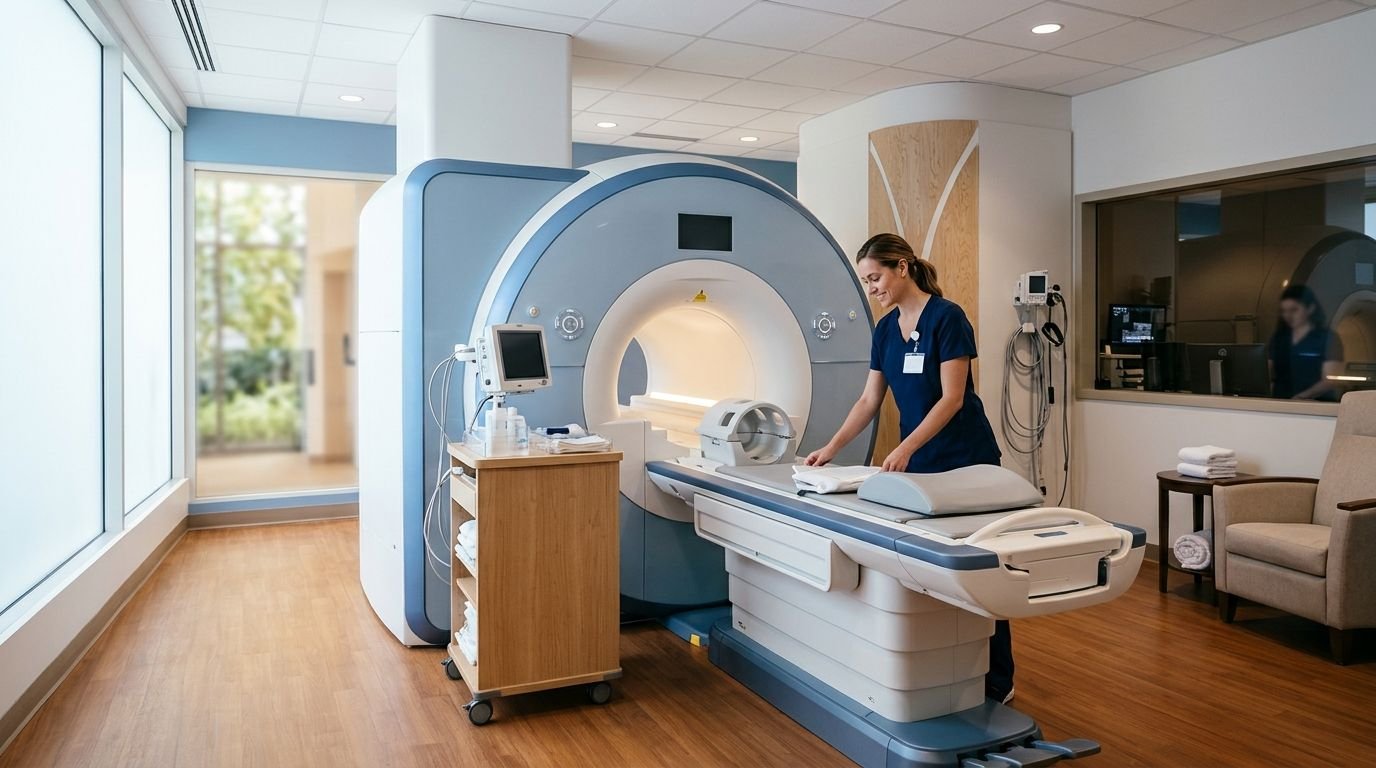
A bone density scan looks at the amount of minerals in bone, whereas an MRI goes deeper into the body’s soft tissue, bone marrow and nerves. Unlike a bone scan for cancer, which uses a radioactive tracer to scan your entire skeleton, an MRI provides high-definition images of specific areas without using radiation, helping the doctor to see if a tumor has spread to your organs, muscles and spine.
MRI is extremely good at imaging soft tissue masses, spinal cord compression, and the brain with exquisite clarity. A cancer bone scan does not even come close when it comes to the level of detail required to properly assess how much disease is present in specific areas. We rely on those images to help make correct diagnoses and plan treatments for our patients in Arizona as well as other states.
3. Can a DEXA Scan Detect Cancer?
No, a DEXA scan measures bone mineral density at the spine and hips to diagnose osteoporosis and assess fracture risk, but it can’t be used to detect tumors or metastases. As per RadiologyInfo.org, the dual-energy X-ray technique isolates true bone density using two low-dose X-ray energies and is therefore not appropriate for the detection of soft tissue masses or cancer.
It uses very low radiation exposure, less than one tenth the amount of a standard chest X-ray, so it prioritizes safety while getting precise bone strength measurements. If cancer in the bones is suspected, another exam is necessary: a bone scan (skeletal scintigraphy) involves using a technetium-99 tracer injected intravenously to look for abnormal bone activity throughout your skeleton. The American Cancer Society says that bone scans are often combined with MRI, PET/CT or biopsy to get a definitive diagnosis. When cancer evaluation is the main objective, MRI comes into play and provides highly detailed images of bone as well as soft tissue.
Why MRI Is Often Preferred for Diagnosis
When we are looking in detail at a specific area, MRI shines because it can produce clear images of both soft tissue and bone marrow. Unlike a bone scan that picks up abnormality on the entire skeleton, MRI concentrates on a specific area with a high level of precision. When doctors want to see something in close-up, MRI is the first choice over a whole body scan.
When it comes to choosing between bone density scan vs. MRI for cancer diagnosis, it really depends on what the clinical question is asking. While bone scan is great for finding if there is whole body bone involvement, it will not be able to give the detail MRI can of one lesion or area that we are interested in. This is why we use both tests – MRI gives the close up while bone scan (or other images) gives us the bigger picture before we get to biopsy and further work up.
The ordering physician ultimately decides which imaging pathway is appropriate for the patient.
5. Bone Scan vs. Bone Density Test
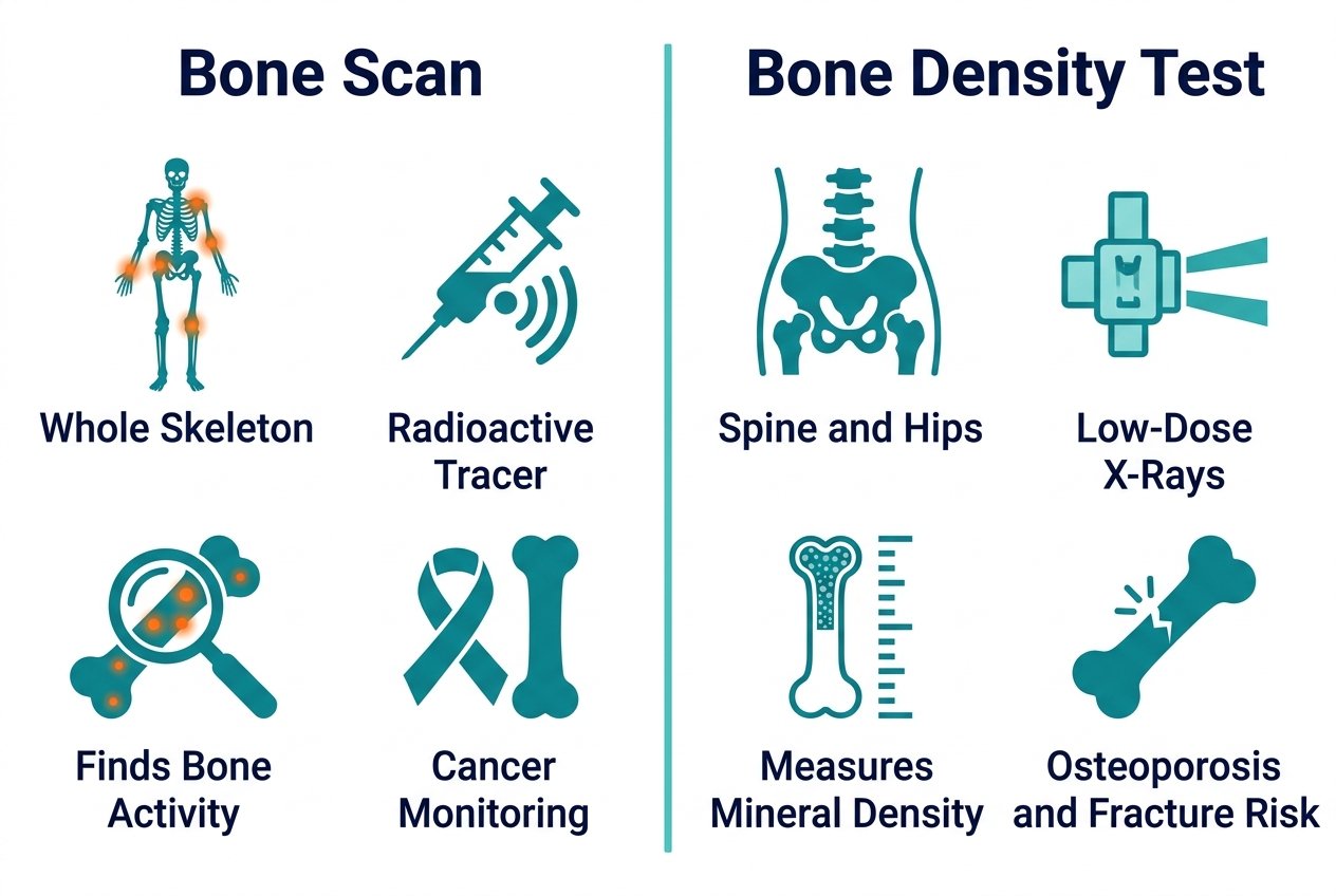
Patients who are researching bone density scan vs MRI for cancer may be unsure if a bone scan and a bone density test are the same exam. No, they are not. A bone scan uses a radioactive tracer to look at all bones in the body for abnormal bone activity, while a bone density test (DEXA scan) looks at bone mineral density at the lumbar spine and hips using dual-energy X-rays.
| Aspect | Bone Scan | Bone Density Test |
|---|---|---|
| Purpose | Detect abnormal bone activity | Measure bone mineral density |
| How it works | Radioactive tracer injection | Dual-energy X-ray beams |
| What it detects | Cancer, fractures, infections | Osteoporosis, fracture risk |
| Body coverage | Entire skeleton | Lumbar spine and hips |
Sometimes a doctor may order both tests to have a more complete view of bone condition.
6. When You Need Both Exams
Many of our patients ask why their doctor orders both a bone scan and a bone density test. These tests are looking at two different things: a bone scan detects any areas where there’s abnormal activity of the bone like metastases, fracture or infection, and a bone density test (DEXA/DXA) is measuring the actual bone mineral density to diagnose if someone has osteoporosis or low bone mass. Being referred to have both doesn’t necessarily mean that you have a problem with your bones; it means that each test is helpful in putting together the complete picture of your bone health.
DEXA scan is the gold standard for diagnosing osteoporosis and evaluating fracture risk. Doctors use T-scores to determine if the results are normal or abnormal: a T-score of -2.5 or lower means osteoporosis, while a score between -1.1 and -2.4 means low bone mass. If you have risk factors such as age, height loss, previous fractures, or long term glucocorticoid use, your doctor may order a DEXA scan along with a bone scan to assess your bone strength as well as any possible abnormalities.
7. Protecting Bones During Cancer Treatment
During cancer treatment, certain therapies can weaken bones, making monitoring essential. We use a bone density scan vs an MRI for cancer-related bone concerns because a DEXA exam is the standard tool for measuring bone mineral density accurately and safely.
For a DEXA scan of the lumbar spine or hip, two low-dose X-ray beams are used to isolate bone mineral density by subtracting soft tissue attenuation. The results are read using T-scores: -2.5 or lower is osteoporosis, while -1.1 to -2.4 indicates low bone mass (osteopenia). Exposure to radiation is minimal, less than one-tenth of that from a chest X-ray, and the outpatient exam takes about 10 to 30 minutes.
After determining that bone density testing is needed, the next step is to schedule the exam with your physician. We collaborate closely with referring teams to make sure the imaging is completed on time, as well as that the results are communicated in an understandable way.
Scheduling Imaging with a Referral
When you receive your referral from your provider, the next step is getting that information turned into an appointment. A good referral will include the exam type, what part of the body to image, and the clinical question, and this information helps speed things along. Ask your referring provider if there are any pre-exam instructions, such as avoiding calcium supplements for 24 hours before your bone density scan or waiting 10–14 days after contrast injections, so we can flag any timing needs when we schedule the exam.
To book, call our scheduling team or use our online portal; we’ll confirm insurance authorization and guide you through any required paperwork. Appointment availability may vary by location and exam type, so we’ll work with you to find a time that fits your schedule. Once booked, you’ll receive a reminder with preparation instructions tailored to your exam.
Frequently Asked Questions
Can a bone density scan detect cancer or bone metastases?
A bone density scan cannot detect cancer or bone metastases. It measures bone mineral density at the spine and hips to diagnose osteoporosis and assess fracture risk using dual-energy X-ray absorptiometry.
Why is an MRI preferred over a bone density scan for cancer diagnosis?
MRI is preferred for cancer diagnosis because it produces clear images of both soft tissue and bone marrow with high precision. Unlike a bone density scan, MRI provides detailed views of specific areas to assess tumor spread or involvement.
What is the difference between a DEXA scan and an MRI for cancer patients?
A DEXA scan measures bone mineral density at the spine and hips to diagnose osteoporosis, while an MRI provides detailed images of soft tissue and bone marrow for cancer evaluation. They address different clinical questions.
Do I need a bone density scan if I am already scheduled for an MRI for cancer?
Whether you need a bone density scan alongside an MRI depends on your doctor’s clinical question. A DEXA scan assesses bone strength and fracture risk, while MRI evaluates cancer involvement in soft tissue and bone marrow.
Why might a doctor order a bone density scan during cancer treatment if it doesn’t detect cancer?
A doctor may order a bone density scan during cancer treatment to monitor bone strength and fracture risk, as certain therapies can weaken bones. The scan measures bone mineral density at the spine and hips using dual-energy X-ray absorptiometry.
Making Sense of Your Imaging Options
Whether to choose a bone density scan or an MRI for cancer depends on what you are trying to find out. The bone density scan (DXA of lumbar spine and hip) is the recommended test when low bone density is suspected, while the bone scan using technetium-99 tracer looks at the whole skeleton to locate areas of increased bone activity that may indicate cancer or fractures.
We support both exam types directly as well as through our provider’s office scheduling. MRI is available in advanced imaging locations throughout Arizona and Texas. Bring your referral and any questions when you come in to schedule. Your provider selected this exam based upon your symptoms, history, and clinical need.
Discuss Your Imaging Options Today
You now understand the key differences between a bone density scan, a bone scan, and an MRI, and how each answers a different clinical question. A DEXA scan measures bone mineral density to assess fracture risk, while MRI provides detailed views of soft tissue and bone marrow when cancer evaluation calls for closer examination. Your referring physician will determine which exam fits your specific situation, since imaging works best as part of a complete diagnostic pathway. If your provider orders an MRI, Arizona Advanced Imaging offers both open and closed MRI at locations across Arizona and Texas, with reports typically returned within 24 to 48 hours. Our team can help coordinate pre-authorization with your insurance plan and answer questions before your appointment. Call (844) 641-2111 to discuss scheduling, or explore our full range of imaging services online. Clear imaging and your physician’s interpretation together give you the information needed for confident next steps.