

Table of Contents
- MRI and Brain Health: Understanding Imaging for Memory Concerns
- What an MRI Can and Cannot Reveal About Alzheimer's
- Early Brain Changes an MRI Can Detect
- Ruling Out Other Causes of Memory Loss
- Brain MRI With or Without Contrast
- Your MRI Experience and Follow-Up Brain Health Monitoring
- Taking the Next Step for Your Brain Health
MRI and Brain Health: Understanding Imaging for Memory Concerns
An MRI for Alzheimer’s and brain health helps evaluate memory concerns by ruling out structural causes like tumors, strokes, or normal-pressure hydrocephalus, but it cannot visualize the microscopic amyloid plaques and tau tangles that define Alzheimer’s disease.
The radiologist looks at the MRIs to check if any other condition is mimicking Alzheimer’s. A normal MRI does not rule out early-stage alzheimer’s, because structural imaging can’t detect the disease’s hallmark biological changes. Around 5 per cent of MRIs done to look for brain change show another underlying treatable cause instead. This is why getting the MRI right is essential in diagnosing dementia.
This article explains what brain imaging can and cannot show when memory concerns arise, how MRI fits into a complete diagnostic workup alongside clinical history and cognitive testing, and how results guide next steps toward clarity and confidence in decision-making.
Key Takeaways
- When your physician orders an MRI for Alzheimer’s and brain health, the scan is designed to answer certain important questions while leaving others unanswered.
- Brain atrophy, in layman terms the loss of brain tissue volume, is an early hallmark of Alzheimer’s-related changes that MRI can visualize before memory symptoms appear.
- An MRI for Alzheimer’s and brain health is only one part of the picture when evaluating memory loss.
- When you arrive for an MRI for Alzheimer’s and brain health monitoring, we want you to feel prepared and at ease.
- Taking an active role in your brain health starts with understanding what an MRI for Alzheimer’s and brain health can reveal — and what it cannot.
What an MRI Can and Cannot Reveal About Alzheimer’s
When your physician orders an MRI for Alzheimer’s and brain health, the scan is designed to answer certain important questions while leaving others unanswered. A normal MRI result does not definitively rule out Alzheimer’s disease because standard imaging cannot visualize the microscopic amyloid plaques and tau tangles that define the condition. Instead, MRI excels at ruling out structural causes of cognitive symptoms—such as tumors, strokes, or normal-pressure hydrocephalus—that may mimic dementia yet often require different treatments.
The real value of MRI, however, rests on its ability to spot other things that can be treated. About 5 percent of imaging scans conducted during the process of screening for dementia come up positive for something the patient was unaware they had until then, although the problem could be treated, as in the case of white matter lesions or past strokes revealed by a scan. Those findings, for example, can point a doctor toward a diagnosis of vascular dementia rather than another type. Also, MRI provides precise three-dimensional measurements of memory structures such as the hippocampus, which supports clinical assessments even though hippocampal shrinkage alone cannot reliably distinguish between dementia types.
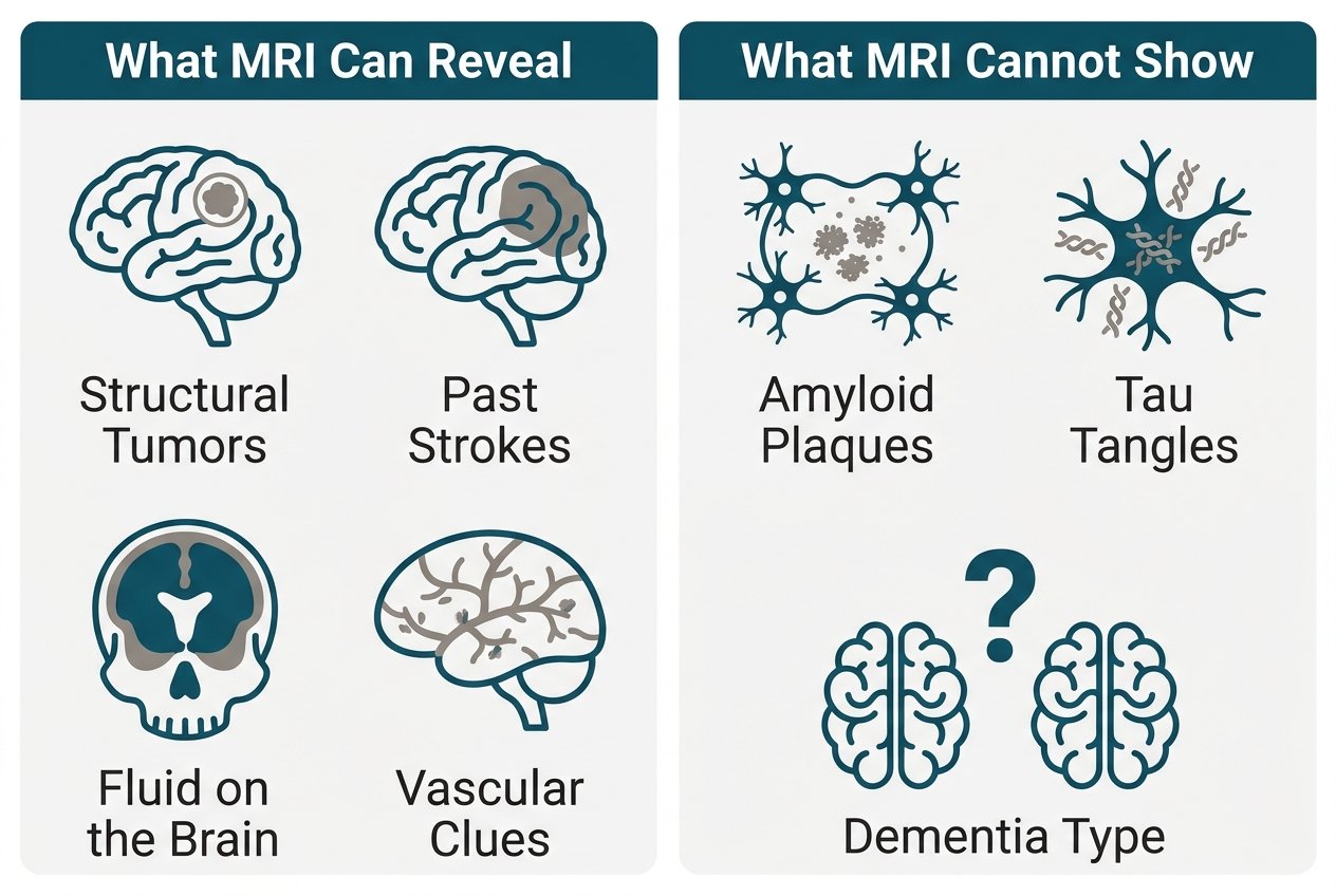
MRI can’t determine all aspects related to Alzheimer’s disease. To make sure whether there are any plaques or tangles in your brain, doctors usually have to order other tests including PET scans, cerebrospinal fluid samples or blood tests. In addition to brain imagery, these additional studies form the entire diagnosis process combined with patients’ medical history and mental state tests as well as lab results. The doctor will coordinate the whole procedure ensuring that each test helps complete the picture of what is going on inside the patient’s head.
Early Brain Changes an MRI Can Detect
Brain atrophy, in layman terms the loss of brain tissue volume, is an early hallmark of Alzheimer’s-related changes that MRI can visualize before memory symptoms appear. Our aim here is to support your health and clarity, and advanced imaging can help identify these subtle shifts years ahead of clinical decline.
A recent study conducted at Johns Hopkins University was published in November 2026 and showed that the entorhinal cortex and amygdala began to shrink five to ten years before the hippocampus, which is normally used to screen people for Alzheimer’s. The entorhinal cortex and amygdala, both important for memory and emotional processing, provide earlier warning signs. This atrophy in these areas correlates strongly with the buildup of tau protein, which disrupts nerve cell function. According to researchers who analyzed data collected through the Alzheimer’s Disease Neuroimaging Initiative along with other related biomarker experiments, monitoring these areas of the brain can allow early-stage testing of anyone between their thirty to forty years old if they have a history of Alzheimer’s in their family.
An MRI for Alzheimer’s and brain health assessment excels at precisely measuring these memory structures in three dimensions, outperforming CT scans in clarity and detail. This capability is why MRI is the preferred imaging modality for evaluating early Alzheimer’s-related atrophy. However, these findings are one piece of a larger puzzle. Your physician uses MRI results alongside your medical history, neurological exams, and lab testing to form a complete picture—never in isolation.
These early changes show why imaging is a critical first step in a thorough evaluation. We need this picture to rule out other potential reasons for memory problems and help us treat those properly in time.
Ruling Out Other Causes of Memory Loss
An MRI for Alzheimer’s and brain health is only one part of the picture when evaluating memory loss. While it can reveal structural changes, a normal scan does not definitively rule out Alzheimer’s disease, because standard MRI cannot visualize the microscopic amyloid plaques and tau tangles that define the condition.
By contrast, its main role is as an exclusion of those causes which mimic dementia but which have treatment (i.e., brain tumour, stroke, subdural haematoma, and normal-pressure hydrocephalus). These account for about 5%, undetected at first sight, of such screening tests. Hence MRI can remain worthwhile even when Alzheimer’s is suspected.
White-matter lesions or past infarcts visible on MRI may suggest vascular dementia rather than Alzheimer’s disease, while hippocampal shrinkage can indicate neurodegeneration but is nonspecific and cannot reliably distinguish between dementia types on its own. Normal-pressure hydrocephalus, in particular, is often mistaken for Alzheimer’s or Parkinson’s disease but may be treatable when identified early through imaging.
A definitive Alzheimer’s diagnosis typically requires additional methods such as PET scans, spinal fluid analyses, or blood biomarkers, and MRI complements rather than replaces a comprehensive workup. However, in general, MRIs complement but don’t replace a thorough evaluation process. In fact, the American Academy of Neurology recommends structural imaging via MRI or noncontrast CT for all patients with dementia symptoms, integrating it with clinical history, neurological examinations, cognitive testing, and laboratory results to form a complete diagnostic picture.
Many folks who get scheduled for MRIs frequently have questions regarding what happens in the procedure. One particular topic is if they’ll require contrast dye.
Brain MRI With or Without Contrast
If there’s any concern about a patient having memory problems, they may need a brain MRI with or without contrast as part of their diagnostic testing, especially if they are suspected to have Alzheimer’s disease or another type of cognitive change. This test allows the radiologists to visualize the brain’s structures in detail, supporting a thorough assessment while helping to rule out treatable conditions that can mimic dementia symptoms. According to the guidelines published by the American Academy of Neurology, structural imaging such as MRI is a standard step in dementia workups and is a natural next step once other potential causes have been considered.
MRI remains generally preferred over CT for memory-loss evaluations because it provides clearer three-dimensional pictures of brain areas involved in memory such as the hippocampus. In comparison to CT, MRI uses magnetic fields instead of radiation and is safer for repeat imaging. Our team follows guidelines that recommend structural imaging as a standard step in dementia workups.
Whether gadolinium contrast should be used depends on the specific clinical question. Contrast enhances blood vessel visualization, but patients should disclose any history of kidney problems or prior adverse reactions to gadolinium in order for us to select the safest option. About 5% of initial studies show abnormalities that have not previously been identified and which require treatment. This emphasizes how radiology is a useful adjunct to both the patient’s medical history and physical exam findings. Findings like white matter lesions or past infarcts may suggest vascular contributions to cognitive decline, guiding the diagnostic path.
Both preparation steps are generally simple and easy. The patient needs to remove metal objects, put on comfortable clothes, and be prepared to lie down for 30-45 minutes. And if sedation was needed due to fear of enclosed places, then it will be necessary for someone else to drive the patient home from the procedure. But really, images aren’t enough on their own-they need to go along with actual neurological exams and laboratory tests in order to create the proper medical diagnosis.
Looking forward to what follows the examination, we will also examine how doctors monitor brain function over time as well as explain what the test means and what you might have to do next.
Your MRI Experience and Follow-Up Brain Health Monitoring
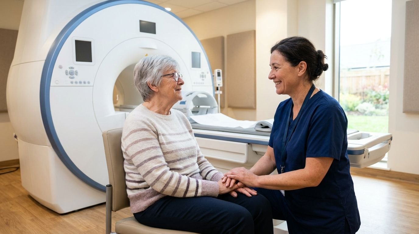
When you arrive for an MRI for Alzheimer’s and brain health monitoring, we want you to feel prepared and at ease. Our team screens every patient for metal objects—jewelry, hairpins, certain makeup—before the scan and will guide you through removing anything that could interfere with the magnetic field. We also ensure you’re comfortable with hearing protection and the stillness required during the exam. MRI technology uses powerful magnets and radio waves, not radiation, so you can safely undergo repeat monitoring exams without concerns about cumulative exposure.
The scan takes around 30 to 45 minutes, depending on what needs to be imaged. You will remain lying still on the table as the machine makes rhythmic humming and tapping sounds. Ear plugs or headphones are given to help protect your ears from these sounds. If you take a sedative for claustrophobia, please make arrangements to have someone drive you home because you won’t be allowed to drive after taking this medication. Our technologist maintains contact using an intercom system and provides a squeezable ball which you may press if you need them to come quickly. During the test, you should be able to hear the technologist’s instruction well enough, but know that they’re always there looking out for your comfort.
Your images will be reviewed by one of our radiologists and a detailed report sent to your physician within 24-48 hours. Your doctor uses this information as part of his/her evaluation of your condition and treatment plan. Imaging provides additional information for the doctor’s diagnosis, but does not replace other medical evaluation. When you come in for a scan at one of our Tucson locations (Tucson or Tucson at 6261 N La Cholla Blvd), remember to check ahead because all of our scanners aren’t available everywhere. Call before coming if you need to make sure you can get the specific type of study that was ordered.
To book or ask questions about your MRI for Alzheimer’s and brain health monitoring, call our centralized scheduling line at 844-641-2111. We’re here to help you take the next step in protecting your brain health over time.
Frequently Asked Questions
Can an MRI definitively diagnose Alzheimer’s disease?
An MRI cannot definitively diagnose Alzheimer’s disease. Standard MRI scans do not visualize the microscopic amyloid plaques and tau tangles that define Alzheimer’s.
What specific changes in the brain can an MRI detect in early Alzheimer’s?
An MRI can detect brain atrophy, particularly shrinkage in the entorhinal cortex and amygdala, which may appear five to ten years before memory symptoms. These changes correlate with tau protein buildup and early Alzheimer’s-related neurodegeneration.
How does an MRI help rule out other causes of memory loss?
An MRI helps rule out structural causes that mimic dementia, such as brain tumors, strokes, subdural hematomas, and normal-pressure hydrocephalus. About 5 percent of MRI scans for memory concerns reveal treatable conditions previously undetected.
What should I expect during an MRI scan for memory evaluation?
During an MRI scan for memory evaluation, you will lie still for 30 to 45 minutes while the machine makes humming and tapping sounds. Ear protection is provided, and you may bring a companion or use sedation if ordered by your physician.
Taking the Next Step for Your Brain Health
Taking an active role in your brain health starts with understanding what an MRI for Alzheimer’s and brain health can reveal — and what it cannot. A brain MRI identifies structural causes behind memory concerns, such as tumors, strokes, or hydrocephalus, but it does not confirm Alzheimer’s disease on its own. A normal scan does not rule out early-stage Alzheimer’s or other dementias, which may require PET imaging, spinal-fluid analysis, or blood biomarkers for a definitive assessment.
Our team partners with your healthcare provider to ensure every MRI is one part of a complete diagnostic workup that includes your medical history, cognitive testing, and lab results.
If you’re concerned about any type of changes in how well your mind works, talk to your doctor to find out what needs to be done. We can help schedule appointments as well as provide information on what patients should know before they get their MRI scan. Results are usually ready within 24-48 hours.
Schedule Your Brain MRI Today
You now understand how a brain MRI fits into a complete Alzheimer’s diagnostic workup — from ruling out treatable causes like tumors, strokes, and normal-pressure hydrocephalus to detecting early structural changes. If your physician has ordered imaging, our team at Arizona Advanced Imaging is ready to help with high-field and Open MRI options. We operate twelve accredited centers across Arizona and Texas, with board-certified, neuro fellowship-trained radiologists reviewing every scan. Reports are typically returned to your referring clinician within 24 to 48 hours, so your next steps are never delayed. Because services and hours vary by location, we encourage you to call ahead and confirm availability at your nearest center. You can reach our centralized scheduling line at (844) 641-2111 for exam questions and appointments. Taking this step for your brain health is simpler than you might think.