

Table of Contents
- The Growing Role of Susceptibility Weighted Imaging in Traumatic Brain Injury
- How Susceptibility Weighted Imaging Works: Physics and Imaging Principles
- SWI for Detecting Microhemorrhages and Axonal Injury in TBI
- Implementing Best‑Practice TBI MRI Protocols with SWI
- Integrating Diffusion Tensor Imaging with SWI for Comprehensive TBI Analysis
- Common Questions About SWI and Advanced TBI Imaging
- Strengthening TBI Diagnosis with SWI and Expert Radiology Partnership
The Growing Role of Susceptibility Weighted Imaging in Traumatic Brain Injury
Beyond conventional sequences, one technique that has gained particular attention is susceptibility weighted imaging (SWI). Its growing role in traumatic brain injury (TBI) stems from superior sensitivity for microhemorrhages and diffuse axonal injury (DAI) that standard sequences often miss. As imaging protocols evolve, SWI is becoming a cornerstone of comprehensive neuroimaging.
SWI offers several clinical advantages for TBI evaluation:
- SWI is 2–4 times more sensitive than conventional gradient-recalled echo (GRE) for detecting hemorrhagic shear injuries, a finding documented in peer-reviewed research from the American Journal of Neuroradiology.
- SWI reveals diffuse axonal injury markers not visible on T2-weighted or FLAIR sequences, improving diagnostic yield.
- Guidelines from the Radiological Society of North America now include SWI in the traumatic brain injury MRI protocol, reflecting its growing standard-of-care status.
- According to the National Institutes of Health, TBI affects millions annually, and SWI can identify chronic microhemorrhages years post-injury, aiding long-term management.
As Your Trusted Radiology Partner, we integrate SWI into our specialized TBI protocols to provide comprehensive evaluations. SWI’s ability to detect microhemorrhages complements other advanced techniques such as diffusion tensor imaging (DTI), which we often incorporate into our exams. Our diagnostic imaging services include advanced TBI protocols that leverage SWI for comprehensive evaluation.
How Susceptibility Weighted Imaging Works: Physics and Imaging Principles
Susceptibility weighted imaging (SWI) is an advanced MRI technique that exploits the magnetic susceptibility differences among tissues to generate high-contrast images. We use T2*-weighted gradient-echo sequences to detect subtle variations in local magnetic fields caused by substances such as deoxyhemoglobin, hemosiderin, ferritin, and calcium deposits. These magnetic perturbations create phase shifts that translate into signal changes, allowing us to visualize venous structures, microbleeds, and calcifications that may be invisible on conventional MRI. According to the American Journal of Neuroradiology, SWI combines magnitude and phase data to enhance sensitivity to these small-field inhomogeneities, making it a powerful tool for neurological assessment.
During image acquisition, we obtain both magnitude and phase images from the T2*-weighted gradient-echo sequence. The magnitude image provides anatomical information, while the phase image reflects local magnetic field variations. We then apply post-processing techniques, including minimum intensity projection (mIP), to suppress background signal and amplify the visibility of small veins and microhemorrhages. Research published by the American Journal of Neuroradiology confirms that combining these steps enhances detection of hemosiderin-laden macrophages, a hallmark of prior microbleeds. The RSNA radiology professional organization provides guidelines on SWI protocols, ensuring standardized image quality across institutions. This multi-step process yields susceptibility-weighted images that significantly improve the detection of pathologies associated with traumatic brain injury (TBI).
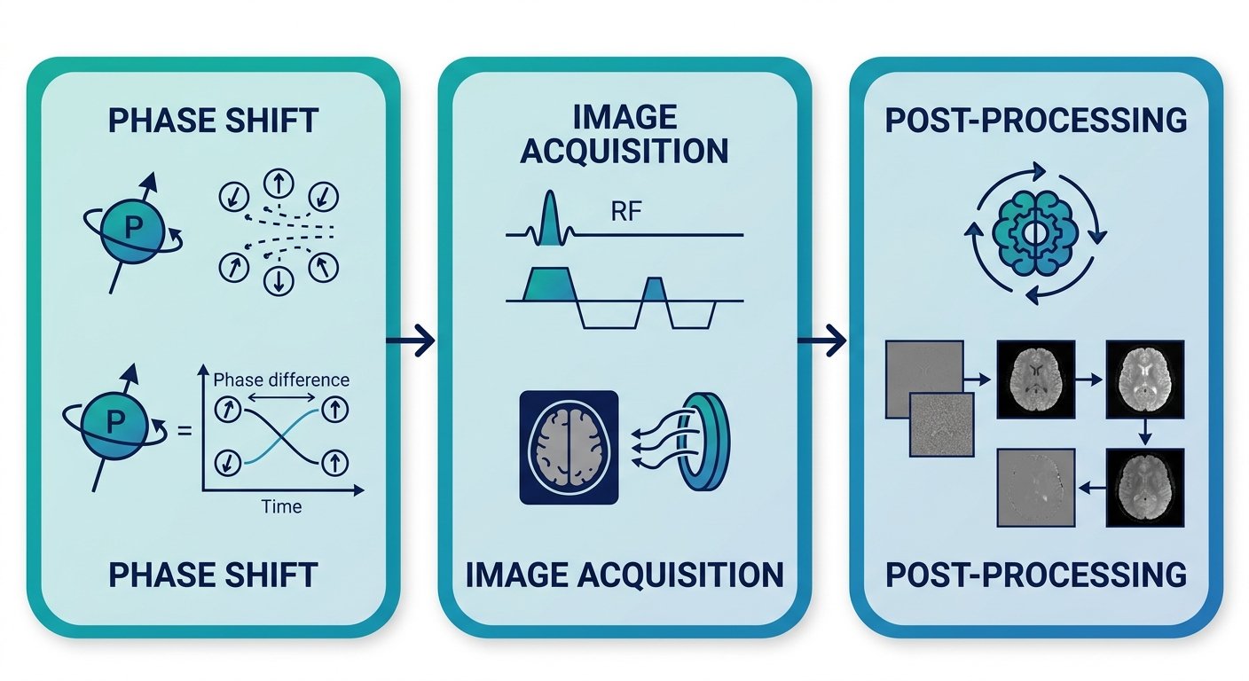
SWI plays a critical role in our traumatic brain injury MRI protocol. It helps detect diffuse axonal injury and microhemorrhages, which are often missed on standard T2-weighted or FLAIR sequences. The National Institutes of Health recognizes SWI’s value in identifying these subtle findings, which can be pivotal in personal injury cases. At Arizona Advanced Imaging, we integrate SWI into every traumatic brain injury MRI protocol across our centers in Phoenix, Arizona and other locations, ensuring that patients and referring physicians benefit from the most advanced neuroimaging techniques available. In addition to TBI assessment, SWI aids in visualizing abnormal venous structures and calcifications in various cerebrovascular conditions. Complementary techniques like diffusion tensor imaging (DTI) are often employed alongside SWI to evaluate white matter tract integrity, providing a comprehensive view of traumatic brain changes. Results may vary based on individual patient conditions.
These principles allow us to detect subtle abnormalities that inform both diagnosis and treatment planning. In the next section, we will explore how these capabilities directly impact patient outcomes in the context of traumatic brain injury care.
SWI for Detecting Microhemorrhages and Axonal Injury in TBI
While T2* GRE can detect some microbleeds, susceptibility weighted imaging (SWI) offers significantly greater sensitivity for identifying cerebral microhemorrhages and diffuse axonal injury after trauma. As part of a comprehensive traumatic brain injury MRI protocol, SWI has become an essential tool for uncovering lesions that conventional sequences may overlook.
The Sensitivity of SWI to Cerebral Microbleeds
SWI exploits the pronounced magnetic susceptibility effects of deoxyhemoglobin and hemosiderin, products of microhemorrhage breakdown. These substances create local magnetic field inhomogeneities that cause signal loss on SWI, allowing even tiny petechial bleeds to be visualized with high contrast. Unlike standard T2-weighted sequences that rely primarily on spin-spin relaxation, SWI enhances contrast by combining magnitude and phase information, making it extraordinarily sensitive to the deposits left by microhemorrhages even weeks or months after injury. This sustained visibility is critical because conventional MRI scans may miss up to 50% of subtle cerebral microbleeds. At Arizona Advanced Imaging, we incorporate SWI into every traumatic brain injury MRI protocol to maximize detection of these subtle lesions and provide objective evidence of trauma that may have significant clinical and legal implications.
SWI vs. T2* Gradient Echo: A Comparison of Microbleed Detection
Comparing SWI with T2* gradient echo (GRE) sequences reveals clear advantages for microbleed identification. The following table summarizes the critical performance differences:
| Feature | SWI | T2* GRE |
|---|---|---|
| Sensitivity to microbleeds | Significantly higher; detects many microbleeds often missed by standard sequences. | Lower sensitivity compared to SWI; limited detection of subtle hemosiderin deposits. |
| Spatial resolution | High-resolution 3D acquisition with fine anatomical detail; phase images aid lesion characterization. | Moderate resolution; images may be degraded by susceptibility artifacts. |
| Contrast mechanism | Exploits magnetic susceptibility effects of deoxyhemoglobin and hemosiderin, providing strong contrast for blood products. | Based on T2* decay from local field inhomogeneities; less specific for microbleeds. |
| Ability to detect small hemorrhages | Excellent; can reliably identify punctate hemorrhages as small as 1–2 mm. | Limited; smaller lesions may fall below detection threshold. |
| Visualization of DAI lesions | Superior for depicting axonal shear injuries, particularly in subcortical white matter and brainstem. | May reveal larger hemorrhagic shears but is less sensitive for non-hemorrhagic or microscopic DAI. |
| Typical acquisition time | Approximately 1–2 minutes per sequence, still within a fast, non-contrast protocol. | Very rapid, often acquired in under one minute. |
The phase image component of SWI enhances detection of paramagnetic substances, while T2* GRE lacks this phase information, leading to reduced conspicuity of small microbleeds. These performance differences are especially important in the setting of mild TBI, where microbleeds may be the only radiologic evidence of injury. Multiple studies published in the AJNR neuroradiology journal have demonstrated that SWI detects significantly more microbleeds than T2* GRE, further supporting its routine use in the assessment of traumatic brain injury. At Arizona Advanced Imaging, SWI is a standard component of our traumatic brain injury MRI protocol, ensuring that no clinically significant microbleed is overlooked.
Identifying Diffuse Axonal Injury with SWI
Diffuse axonal injury (DAI) results from rotational acceleration-deceleration forces that shear white matter tracts, producing microscopic lesions that are often invisible on CT and conventional MRI. SWI is uniquely sensitive to the hemorrhagic component of DAI because the associated microbleeds contain deoxyhemoglobin and hemosiderin deposits. These lesions characteristically appear as punctate hypointensities at the grey–white matter junction, in the corpus callosum, and in the brainstem. When combined with diffusion tensor imaging (DTI), which assesses white matter integrity, SWI provides a more comprehensive picture of axonal injury. At Arizona Advanced Imaging, we use the CBI-M framework to standardize the classification of DAI based on SWI findings, ensuring that even subtle microbleeds are documented and correlated with clinical severity. In many cases, the microhemorrhages revealed by SWI are the only objective imaging evidence of trauma, making this sequence indispensable in personal injury evaluations. Our experienced neuroradiologists carefully review SWI images to detect these telltale signs of axonal shear, which can explain persistent neurological symptoms even when other MRI sequences appear normal.
Results may vary based on individual patient conditions. Please consult with a healthcare professional for diagnosis and treatment advice.
Implementing Best-Practice TBI MRI Protocols with SWI
Given the limitations of conventional MRI for mild TBI, implementing specialized protocols becomes essential. The addition of advanced sequences like susceptibility weighted imaging swi transforms our ability to detect subtle hemorrhagic and axonal injuries that standard approaches often miss. Our centers integrate these refined techniques into every dedicated head injury examination to produce clearer answers for referring physicians, patients, and legal teams.
Standard MRI Sequences for Traumatic Brain Injury Assessment
A well-designed traumatic brain injury MRI protocol begins with five core sequences that together survey the brain for structural abnormalities. Each sequence contributes unique diagnostic information and cannot be omitted without risking incomplete evaluation.
T2-weighted imaging provides high-resolution anatomical detail, revealing contusions, edema, and mass effect. FLAIR suppresses cerebrospinal fluid signal to highlight periventricular and cortical lesions that T2 alone may obscure. T2-weighted gradient echo* detects blood products such as hemosiderin deposits from prior hemorrhage. Susceptibility weighted imaging serves as the most sensitive tool for identifying microhemorrhages and venous abnormalities. Finally, diffusion weighted imaging captures acute ischemic changes and cellular swelling that accompany traumatic axonal injury.
Together, these sequences form the foundation of a reliable TBI examination. When acquisition parameters are optimized—such as obtaining SWI with whole-brain coverage and slice thickness ≤3 mm—the protocol delivers the resolution needed to identify abnormalities that influence both clinical management and legal determinations.
Incorporating SWI into TBI Protocols: Guidelines and Recommendations
We follow a structured approach when integrating susceptibility weighted imaging swi into every dedicated brain trauma study. The 2026 AAN/CDC guidelines recommend SWI over standard GRE for detecting hemorrhagic shear injuries, and our workflow reflects that consensus. In practice, we combine SWI with diffusion tensor imaging dti when the clinical question involves suspected diffuse axonal injury.
The table below contrasts the elements of a minimal TBI protocol against the expanded version we employ as part of the spintech MRI TBI protocol. This standardized framework incorporates SWI, DTI, and quantitative susceptibility mapping for a more complete picture of post-traumatic pathology.
| Sequence | Standard TBI Protocol | Advanced TBI Protocol (with SWI/DTI) |
|---|---|---|
| T2-weighted | Axial, 4–5 mm slice thickness; screens for mass effect and contusions | High-resolution axial and coronal planes for detailed anatomical mapping |
| FLAIR | Axial, 4–5 mm; identifies periventricular and subcortical lesions | 3D acquisition with ≤3 mm slices improves subtle lesion conspicuity |
| Gradient Echo | Axial, 4–5 mm; detects large hemorrhages | Replaced or supplemented by SWI for increased sensitivity |
| SWI | May be omitted in minimal protocols | Whole-brain coverage, ≤3 mm slice thickness; enhanced detection of microhemorrhages and venous injury |
| DWI | Axial, b=1000; screens for ischemia and cytotoxic edema | Maintained with high b-value for acute injury evaluation |
| DTI | Not included | Added with fractional anisotropy mapping and tractography to evaluate diffuse axonal injury |
The inclusion of diffusion tensor imaging dti moves the study beyond standard structural assessment. By measuring fractional anisotropy and generating tractography maps, we can identify disruptions in white matter integrity that correlate with cognitive and functional deficits. These findings are particularly relevant in personal-injury cases where objective evidence of injury supports the clinical narrative.
For consistent results, every study adheres to acquisition parameters recognized by professional radiology organizations. The Radiological Society of North America provides technical standards that guide our sequence selection and quality assurance practices.
Report Turnaround and Fellowship Training: Ensuring Clinical and Legal Utility
Even the most technically precise images lose value without timely, expert interpretation. Our 24 – 48 Hour Report Turnaround standard developed from the practical reality that referring physicians need answers to make treatment decisions and attorneys require documentation before depositions. Delayed reports stall patient care and weaken litigation support, which is why we prioritize rapid delivery without sacrificing accuracy.
Fellowship-trained neuroradiologists bring specialized pattern-recognition skills that directly impact diagnostic confidence. Their training focuses on the types of subtle findings—punctate microhemorrhages on SWI, mild anisotropy reductions on DTI—that general radiologists may overlook or dismiss as clinically insignificant. When board-certified neuroradiologists review these studies, the interpretation benefits from years of concentrated experience with the very conditions that TBI protocols are optimized for detecting.
We invest in this expertise because the consequences of missed findings ripple through both clinical and legal pathways. A patient with undetected axonal injury may not receive appropriate rehabilitation, and a legitimate injury claim may fail without imaging evidence. Rapid turnaround reinforces that early insight, giving stakeholders the information they need within the timeframes that matter.
With a reliable protocol in place, the next step is ensuring timely, expert interpretation to support both clinical decision-making and personal-injury litigation.
Integrating Diffusion Tensor Imaging with SWI for Comprehensive TBI Analysis
At Arizona Advanced Imaging, our traumatic brain injury MRI protocol integrates susceptibility weighted imaging (SWI) with diffusion tensor imaging (DTI) to capture the full spectrum of post-traumatic pathology. SWI excels at detecting microhemorrhages and shearing injuries, while DTI reveals white-matter tract disruption—together they form a more complete injury profile than either sequence alone.
While DTI and SWI each reveal different aspects of TBI pathology, their integration offers a more complete picture. SWI is highly sensitive for hemorrhagic contusions, shear injuries, and microbleeds that conventional GRE sequences often miss. In practice, we acquire high-resolution SWI with optimized echo times to improve visualization of tiny hemosiderin deposits that standard sequences may miss. As peer-reviewed research in the American Journal of Neuroradiology confirms, SWI’s blood-iron detection capability makes it indispensable for identifying diffuse vascular injury. In our protocols, this susceptibility weighted imaging SWI capability uncovers subtle hemorrhage that can correlate with cognitive and functional deficits.
DTI complements SWI by measuring fractional anisotropy (FA) and mean diffusivity (MD) to quantify axonal integrity. Where SWI identifies bleeding, diffusion tensor imaging DTI maps non-hemorrhagic white-matter disconnection—capturing damage that blood-sensitive sequences cannot show. We routinely pair quantitative DTI metrics and tractography with SWI findings to localize disrupted pathways and produce structured imaging summaries that support early multidisciplinary care and rehabilitation planning. The National Institutes of Health and the Society of Nuclear Medicine and Molecular Imaging have highlighted that integrating these biomarkers improves TBI severity grading and prognosis, helping us identify patients who may benefit from early intervention.
Because cardiovascular comorbidities can influence recovery trajectories—as highlighted in recent AHA journals cardiovascular research—we ensure our TBI assessments account for these systemic factors, delivering Clarity in every image, confidence in every decision.
Common Questions About SWI and Advanced TBI Imaging
Q: What is susceptibility weighted imaging (SWI) and how does it help diagnose TBI in Phoenix? A: SWI is an advanced MRI sequence sensitive to microhemorrhages. It detects diffuse axonal injury often missed by standard scans, helping diagnose TBI in Phoenix.
Q: What does a traumatic brain injury MRI protocol include? A: Our TBI protocol includes susceptibility weighted imaging (SWI), diffusion tensor imaging dti, and conventional T1/T2/FLAIR sequences to assess structural and microstructural damage.
Q: How does DTI complement SWI in evaluating white matter? A: Diffusion tensor imaging maps white matter tract integrity, identifying axonal injury, while SWI pinpoints blood products. Together they offer a comprehensive view of traumatic brain injury.
Q: Why is advanced imaging important for Arizona personal injury claims? A: Our CBI-M framework integrates SWI and DTI findings to objectively grade injury severity. Court-ready reports support legal documentation, and we offer lien-based billing and 24–48 hour turnaround for Arizona patients.
Please consult your healthcare professional for medical diagnosis and treatment advice.
Strengthening TBI Diagnosis with SWI and Expert Radiology Partnership
Susceptibility weighted imaging (SWI) significantly strengthens traumatic brain injury diagnosis by detecting microhemorrhages that standard MRI may miss.
Our expert radiologists, trained in specialized traumatic brain injury MRI protocols, interpret SWI results alongside complementary techniques like diffusion tensor imaging (DTI) to produce court-ready reports. This nuanced approach reduces the risk of overlooked subtle injuries and supports personal injury cases.
We offer SWI across our Arizona and Texas locations with a 24–48 hour report turnaround, backed by our full suite of diagnostic imaging services in Arizona. Your health and clarity are our priority. Please consult with a healthcare professional for diagnosis and treatment advice.